Why NanoZoomer
Products
Solutions
Case study
Resources


Japan (EN)
Select your region or country.
A revolution in histopathology education
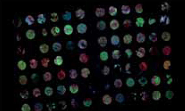
Whole Slide Imaging (WSI) scanner also known as virtual microscopy is rapidly proving to be a useful tool in research and academia. Histopathological preparations are digitalized and can be shared simultaneously across an entire classroom of students. Using those shared information facilitates interactions with the teacher and among students, directly improving their ability to learn. This technology transforms the educational process by enabling the possibility to screen share but also to mark or annotate and more. Universities are making the change from traditional microscopes to implement this revolutionary way of teaching, especially in the field of histopathology education.
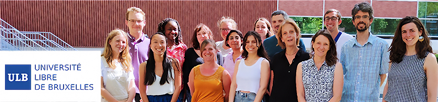
These teaching transitions vary depending on the educational situation and often need support. The teaching of histology at Université Libre de Bruxelles (ULB) is delivered to around 800 students of various disciplines (Medicine, veterinary medicine, dentistry, biomedical sciences, physiotherapy, …). The implementation of virtual microscopy in the Faculty of Medicine at ULB was a step-by-step process over time, which ended with their whole equipment being installed in-house.
For a number of years, the histology teachers relied on an external facility who provided them with scanned slides, used for their teaching and sometimes research. These slides were first converted into tiles by an in-house solution, and then added to their online learning platform Moodle, where they were visualized through the application Zoomify. However, this process was far from ideal based on the reliance of using an external facility. In addition, the teachers wished to further increase their flexibility concerning image sizing, storage capacity, user’s rights and function availability. That is why they sought another solution in association with the Moodle platform. By doing so, they hoped to achieve a smoother process to save time in class preparations.
The right combination between hardware and software
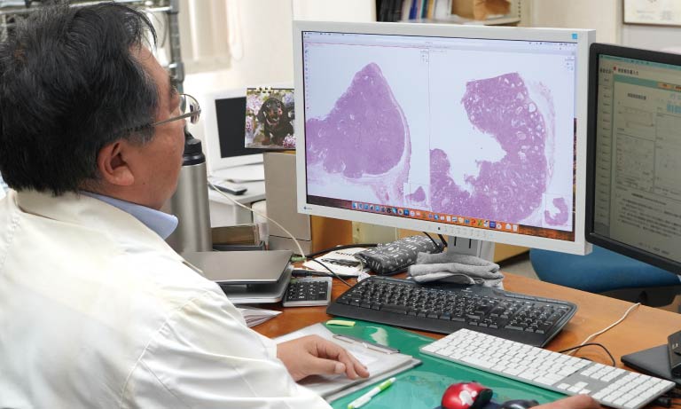
Pascale Lybaert, DVM, PhD, responsible for histology course in the Veterinary Medicine curriculum sought a cost efficient, smoother and more versatile workflow to improve the autonomous work from students to support the theoretical approach given ex-cathedra to vet students. Dr. Lybaert knew Hamamatsu Photonics’ NanoZoomer whole slide scanners well and was confident in the quality of the digital images for her classes. Combined with the right software, this solution would alleviate some of her unnecessary time-consuming workload.
The appearance of the COVID-19 pandemic greatly accelerated the requirement for an updated integrative workflow as very suddenly, universities were forced to teach at a distance. The educational field had to adapt quickly to continue providing classes for their students. Microscopes were no longer an option in this situation, digital scanners however, proved ideal for distance learning. Since the preparation is digitalized completely, it can be shared instantly and can replace the in-person microscopic observation, allowing all classes to continue undisrupted from home.
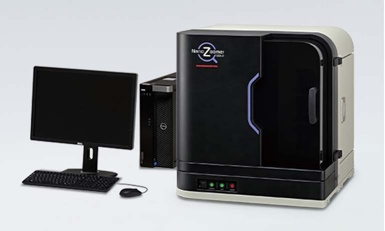
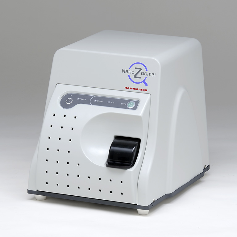
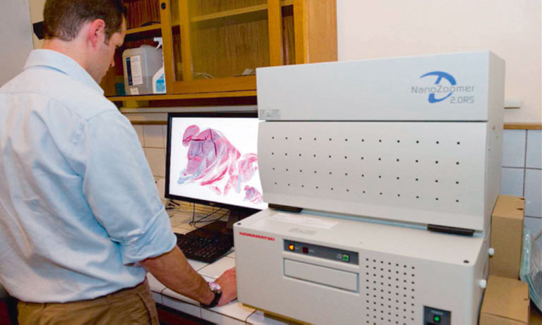
Therefore, Dr. Lybaert approached Hamamatsu Photonics and ended-up picking from the whole slide scanners series, the NanoZoomer®-SQ , which best suited her requirements. She enjoyed the benefit of being able to do all of her work from her desk. The NanoZoomer®-SQ is compact and easy to use wherever you need it to be. As she liked reviewing each slide before her classes, this solution allowed her to do this, right from the comfort of her desk.
"Thanks to this new solution, I was able to divide my class into two groups, one remote and one on microscopes. This meant that I could spend quality time with a smaller group of students. It makes a real difference for all of us.”

Pascale Lybaert, DVM, PhD
Responsible for histology course in the Veterinary Medicine curriculum
Université Libre de Bruxelles (ULB)
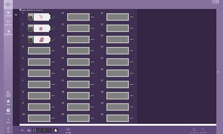
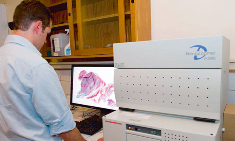
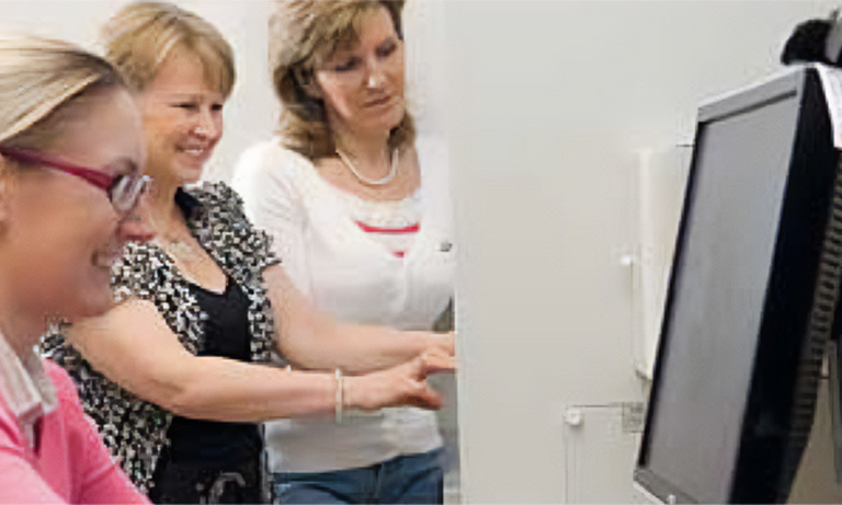
She placed each glass slide into the device to scan and through the dedicated NZConnect software, she was able to easily create, view and perform quality-checks on whole slide images from her computer. She decided to keep the microscopes for the more practical sessions, when students were able to be present physically, and use the digital scanner for distance learning, practical preparation and homework. This gave her the freedom to approach her teaching methods differently. For example, it allowed her to divide the classroom into groups so she could focus her time on smaller groups of students, enhancing the quality of the teaching experience.
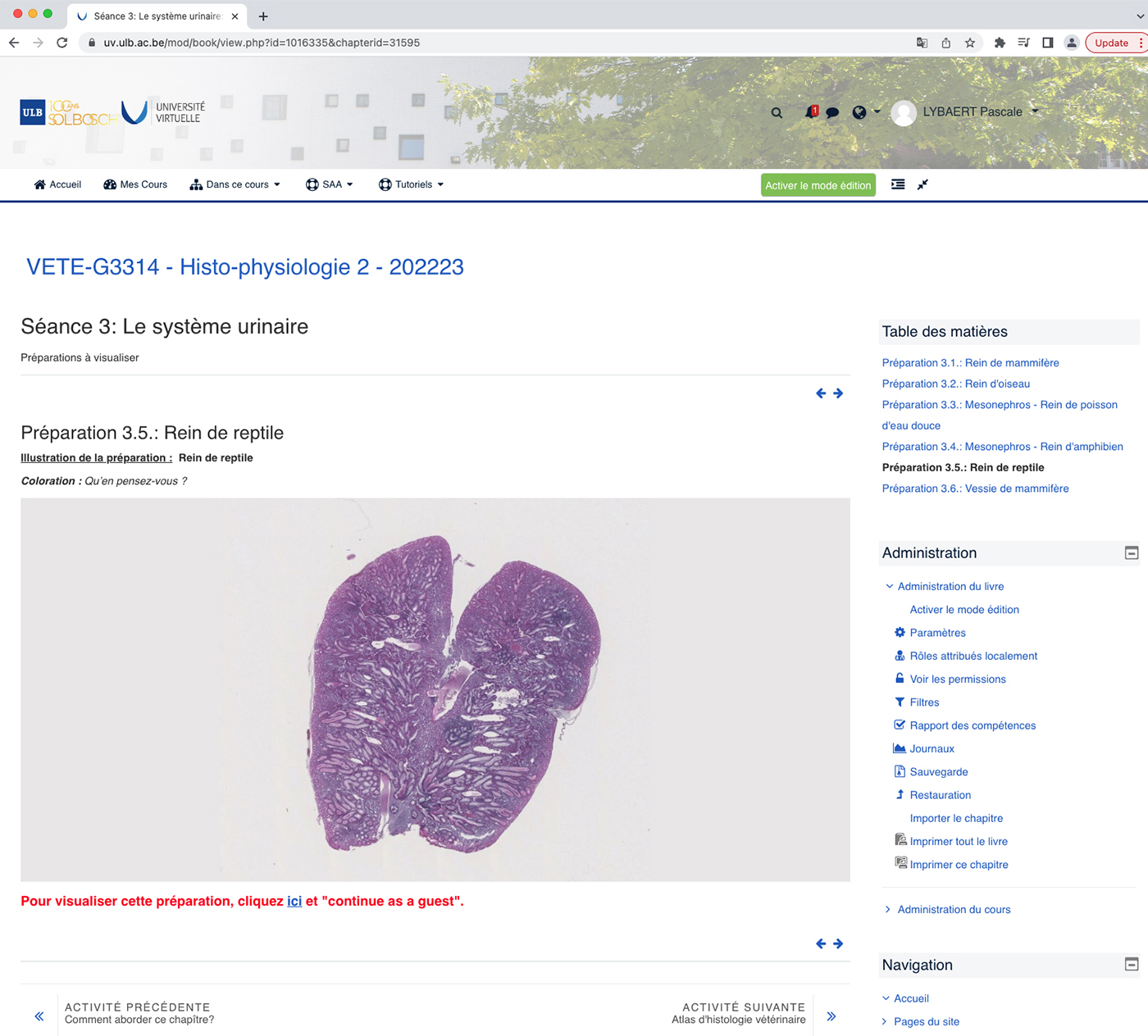
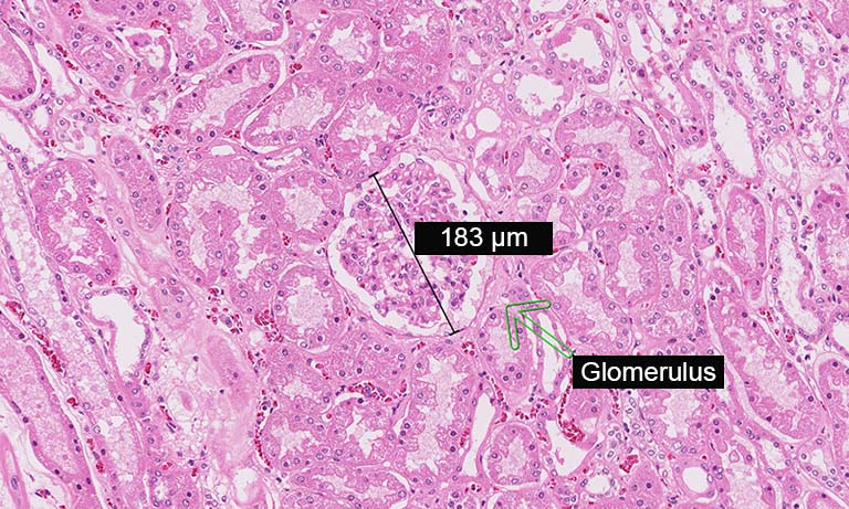
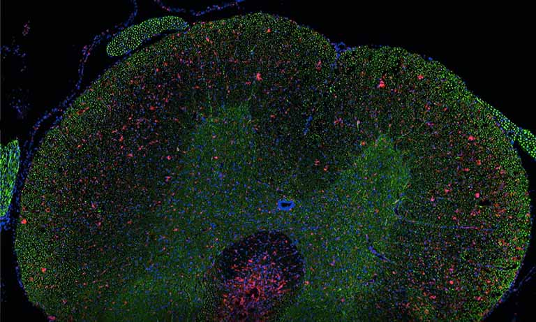
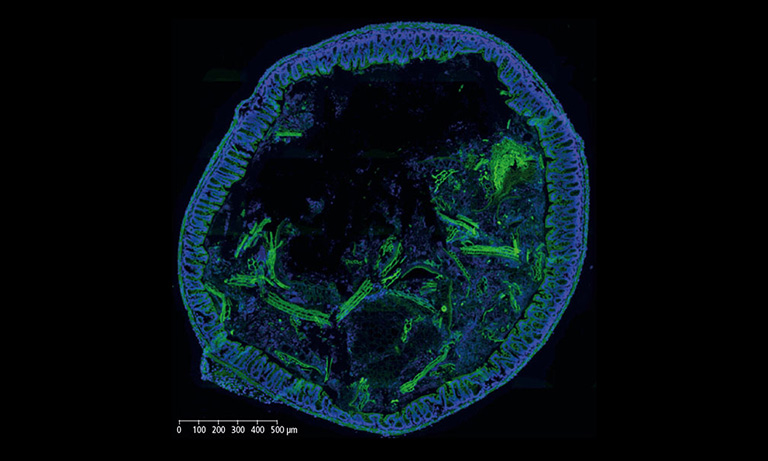
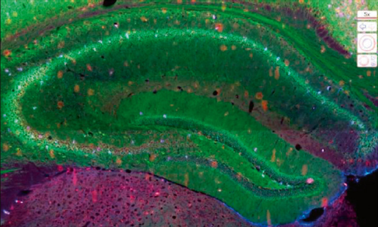
On screen visual of a reptile kidney from NanoZoomer®-SQ scan
This image viewing software combined well with their online teaching management tool, Moodle. It was the perfect extension to manage the digital images for her and her student’s needs. From their computers, the student were able to view and click on the images, add annotations, make measurements etc. This meant that for example, she was able to conduct exams from a distance while ensuring the highest quality data. Finally, as the software is available on PC and Mac and, the number of digital slides stored is unlimited, this made it a flexible and sustainable option for teaching but also research in academia. Indeed, the use of the NanoZoomer®-SQ was rapidly extended to the digitalization of the histological preparation realized in several research projects of the Research Laboratory on Human Reproduction (lab host of the NanoZoomer®-SQ) but also a number of other research labs of the ULB Faculty of Medicine. Digitalization allows an effective and comfortable post-examination and analysis of the preparation as well as unlimited storage of those data.
Many universities around the world are transitioning to digital microscopy as a new educative technology to stimulate the learning experience. Easy to use and interactive, it allows for a more collaborative approach and supports a different way of learning, especially for histopathology students.
The NanoZoomer team at Hamamatsu Photonics is delighted to be able to contribute to supporting histopathology research studies in the hope of finding better diagnosis for patients around the world.
About the Research Laboratory on Human Reproduction
Created by Professor Yvon Englert in 1998, the Research Laboratory on Human Reproduction aimed to improve our knowledge on different aspect of male and female reproduction and to improve the diagnosis and treatment of infertility.
During her PhD in the lab, Isabelle Demeestere has pioneered the field of fertility preservation in women during cancer treaments by the cryopreservation of ovarian tissue. She became head of the laboratory in 2016. The research projects of the lab have been focused mainly on the development of innovative approaches to develop fertility preservation strategies and to expand our understanding of male and female reproductive function. Professor Pascale Lybaert joined the lab in 2018 to bring her expertise in male reproduction to expand the scope of fertility preservation to the male partner.
The lab has pioneered in the development of fertility preservation techniques and works on different approaches to improve those techniques in children and young men and women at risk of future infertility due to their diseases (such as genetic or gynecological disorders) or to oncological treatments (such as chemotherapy and radiotherapy). The lab was able to be amongst the earliest innovators in this field thanks to its close collaboration with the Fertility Clinic at Erasme Hospital, and with a multidisciplinary team including technicians, data manager, medical gynecologists, oncologists, and scientists.
Thanks to this expertise, the lab joined the ULB-Research cancer center (U-CRC) that gathers over 190 scientists and physicians from 19 groups working on various aspects of cancer research.

Other research case study
- Confirmation
-
It looks like you're in the . If this is not your location, please select the correct region or country below.
You're headed to Hamamatsu Photonics website for JP (English). If you want to view an other country's site, the optimized information will be provided by selecting options below.
In order to use this website comfortably, we use cookies. For cookie details please see our cookie policy.
- Cookie Policy
-
This website or its third-party tools use cookies, which are necessary to its functioning and required to achieve the purposes illustrated in this cookie policy. By closing the cookie warning banner, scrolling the page, clicking a link or continuing to browse otherwise, you agree to the use of cookies.
Hamamatsu uses cookies in order to enhance your experience on our website and ensure that our website functions.
You can visit this page at any time to learn more about cookies, get the most up to date information on how we use cookies and manage your cookie settings. We will not use cookies for any purpose other than the ones stated, but please note that we reserve the right to update our cookies.
1. What are cookies?
For modern websites to work according to visitor’s expectations, they need to collect certain basic information about visitors. To do this, a site will create small text files which are placed on visitor’s devices (computer or mobile) - these files are known as cookies when you access a website. Cookies are used in order to make websites function and work efficiently. Cookies are uniquely assigned to each visitor and can only be read by a web server in the domain that issued the cookie to the visitor. Cookies cannot be used to run programs or deliver viruses to a visitor’s device.
Cookies do various jobs which make the visitor’s experience of the internet much smoother and more interactive. For instance, cookies are used to remember the visitor’s preferences on sites they visit often, to remember language preference and to help navigate between pages more efficiently. Much, though not all, of the data collected is anonymous, though some of it is designed to detect browsing patterns and approximate geographical location to improve the visitor experience.
Certain type of cookies may require the data subject’s consent before storing them on the computer.
2. What are the different types of cookies?
This website uses two types of cookies:
- First party cookies. For our website, the first party cookies are controlled and maintained by Hamamatsu. No other parties have access to these cookies.
- Third party cookies. These cookies are implemented by organizations outside Hamamatsu. We do not have access to the data in these cookies, but we use these cookies to improve the overall website experience.
3. How do we use cookies?
This website uses cookies for following purposes:
- Certain cookies are necessary for our website to function. These are strictly necessary cookies and are required to enable website access, support navigation or provide relevant content. These cookies direct you to the correct region or country, and support security and ecommerce. Strictly necessary cookies also enforce your privacy preferences. Without these strictly necessary cookies, much of our website will not function.
- Analytics cookies are used to track website usage. This data enables us to improve our website usability, performance and website administration. In our analytics cookies, we do not store any personal identifying information.
- Functionality cookies. These are used to recognize you when you return to our website. This enables us to personalize our content for you, greet you by name and remember your preferences (for example, your choice of language or region).
- These cookies record your visit to our website, the pages you have visited and the links you have followed. We will use this information to make our website and the advertising displayed on it more relevant to your interests. We may also share this information with third parties for this purpose.
Cookies help us help you. Through the use of cookies, we learn what is important to our visitors and we develop and enhance website content and functionality to support your experience. Much of our website can be accessed if cookies are disabled, however certain website functions may not work. And, we believe your current and future visits will be enhanced if cookies are enabled.
4. Which cookies do we use?
There are two ways to manage cookie preferences.
- You can set your cookie preferences on your device or in your browser.
- You can set your cookie preferences at the website level.
If you don’t want to receive cookies, you can modify your browser so that it notifies you when cookies are sent to it or you can refuse cookies altogether. You can also delete cookies that have already been set.
If you wish to restrict or block web browser cookies which are set on your device then you can do this through your browser settings; the Help function within your browser should tell you how. Alternatively, you may wish to visit www.aboutcookies.org, which contains comprehensive information on how to do this on a wide variety of desktop browsers.
5. What are Internet tags and how do we use them with cookies?
Occasionally, we may use internet tags (also known as action tags, single-pixel GIFs, clear GIFs, invisible GIFs and 1-by-1 GIFs) at this site and may deploy these tags/cookies through a third-party advertising partner or a web analytical service partner which may be located and store the respective information (including your IP-address) in a foreign country. These tags/cookies are placed on both online advertisements that bring users to this site and on different pages of this site. We use this technology to measure the visitors' responses to our sites and the effectiveness of our advertising campaigns (including how many times a page is opened and which information is consulted) as well as to evaluate your use of this website. The third-party partner or the web analytical service partner may be able to collect data about visitors to our and other sites because of these internet tags/cookies, may compose reports regarding the website’s activity for us and may provide further services which are related to the use of the website and the internet. They may provide such information to other parties if there is a legal requirement that they do so, or if they hire the other parties to process information on their behalf.
If you would like more information about web tags and cookies associated with on-line advertising or to opt-out of third-party collection of this information, please visit the Network Advertising Initiative website http://www.networkadvertising.org.
6. Analytics and Advertisement Cookies
We use third-party cookies (such as Google Analytics) to track visitors on our website, to get reports about how visitors use the website and to inform, optimize and serve ads based on someone's past visits to our website.
You may opt-out of Google Analytics cookies by the websites provided by Google:
https://tools.google.com/dlpage/gaoptout?hl=en
As provided in this Privacy Policy (Article 5), you can learn more about opt-out cookies by the website provided by Network Advertising Initiative:
http://www.networkadvertising.org
We inform you that in such case you will not be able to wholly use all functions of our website.
Close