Why NanoZoomer

Products

Solutions

Case study
Resources


United Kingdom (EN)
Select your region or country.
Digital Veterinary Pathology Workflow
Published on March 12, 2025
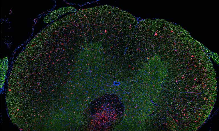
Digital Veterinary Pathology Test
Amanecer Co., Ltd. has been digitizing veterinary samples using whole slide scanners since 2013 and adopted the NanoZoomer in 2020. Their goal is to enhance test quality and service efficiency through a digital pathology workflow.

Amanecer Co., Ltd.
Headquartered in its own building in Sapporo, Hokkaido. Amanecer Co., Ltd. handles approximately 30 000 pathology test requests annually (as of 2024) from over 6000 contracted veterinary clinics across Japan, supported by their offices in Tokyo and Osaka.
Digital veterinary pathology workflow of Amanecer Co., Ltd.
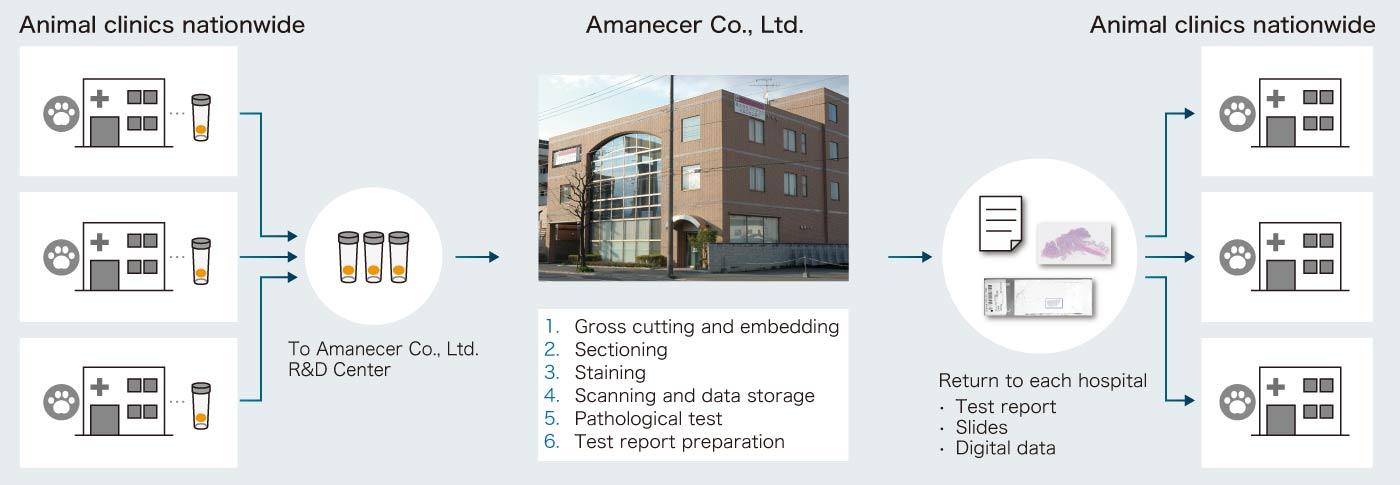
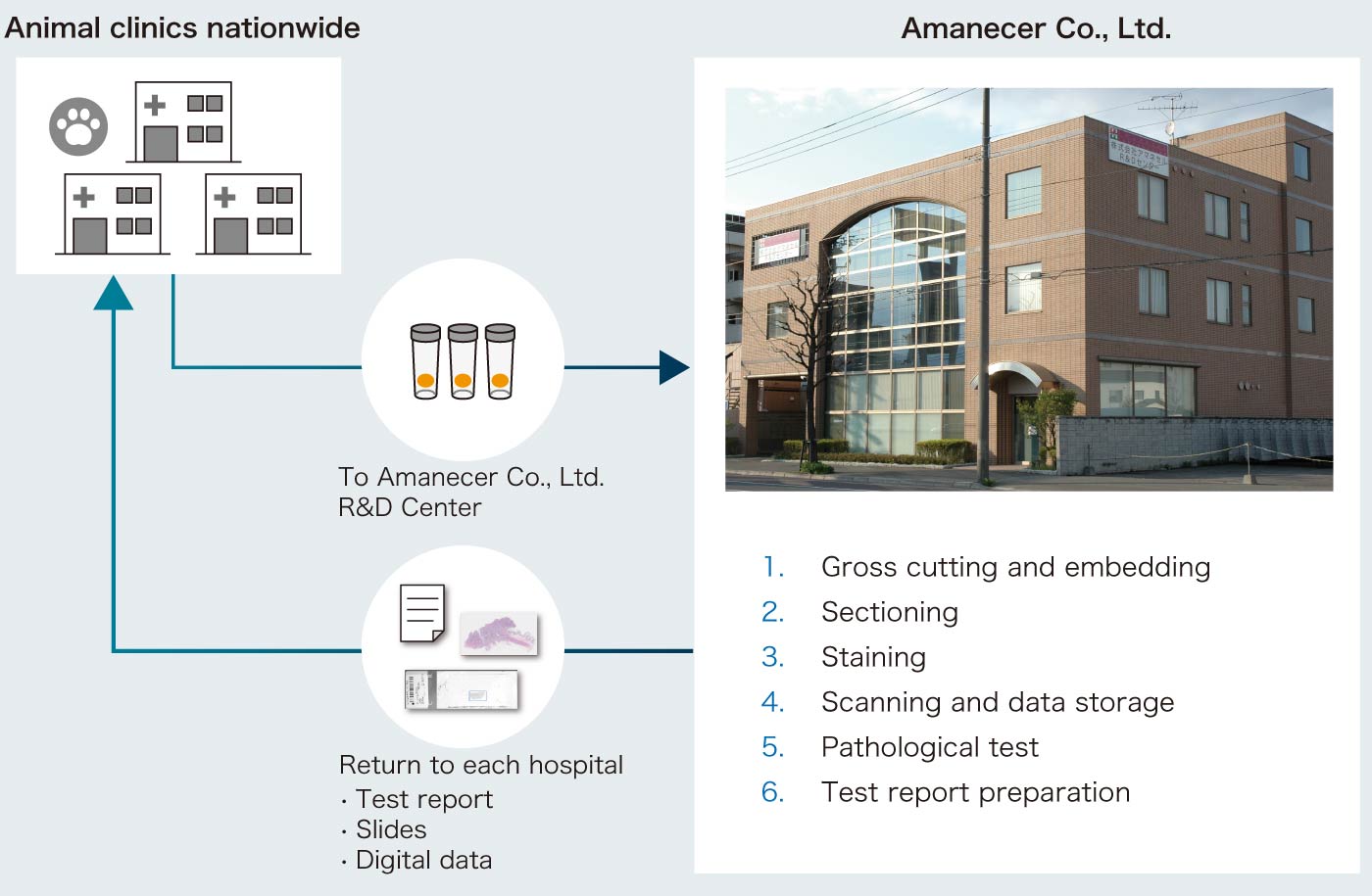
A workflow in Amanecer Co., Ltd
1. Gross cutting and embedding
2. Sectioning
The wet organs we receive are cut as needed and then processed into paraffin-embedded blocks.
To accommodate the unique characteristics of each tissue, sectioning is performed manually using one of eight microtomes.


3. Staining
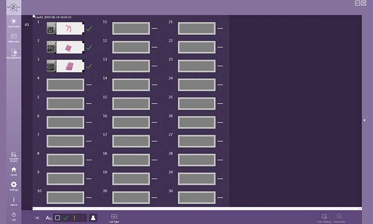
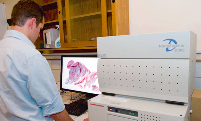
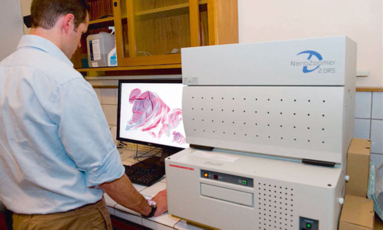
4. Scanning and Data Storage
(H&E Staining/Special Staining/Immunostaining)
The wet organs we receive are cut as needed and then processed into paraffin-embedded blocks.
The wet organs we receive are cut as needed and then processed into paraffin-embedded blocks.


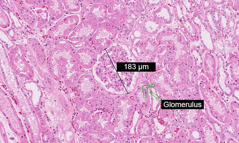
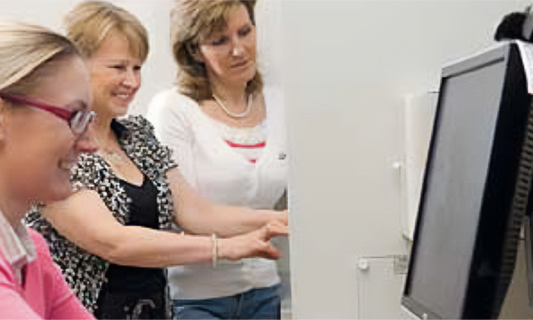
5. Pathological Test
6. Test Report Preparation
(Vet pathologists)
We guarantee highly accurate tests based on over 20 years of experience in veterinary pathology and annual record of about 30 000 cases.
We create test reports using captured images from either a whole slide scanner or a microscope.


Digitalization of veterinary pathology
Q1: How do you use digital pathology for veterinary purposes?
Our general workflow involves preparing slides from samples sent by animal clinics nationwide, conducting tests, and submitting reports. We use whole slide scanners to digitize slides, enabling us to test and capture images for test reports. Currently, we digitize about half of our pathology tests (approximately 50 cases per day). Ideally, we would like to scan all slides digitally, but certain samples are better tested using a microscope. Therefore, we use a combination of digital and traditional methods. We believe this hybrid approach allows us to deliver more accurate tests and better services.
Q2: Why did you implement a digital pathology workflow?
Our primary motivation for adopting digital pathology was to enable remote tests from outside the office. Additionally, we wanted to embrace innovation as a veterinary company. We introduced whole slide scanners in 2013 and began implementing a digital pathology workflow. Although we faced initial challenges in scanning slides during the first few months, we were able to overcome them and have since been consistently delivering reliable pathology services.

Q3: What are the benefits of digitalization?
Before implementing digital pathology, we stored all slides physically and had to retrieve them manually from our warehouse when needed, even for cases tested a month prior. This retrieval process was time-consuming and often added significantly to our workload.
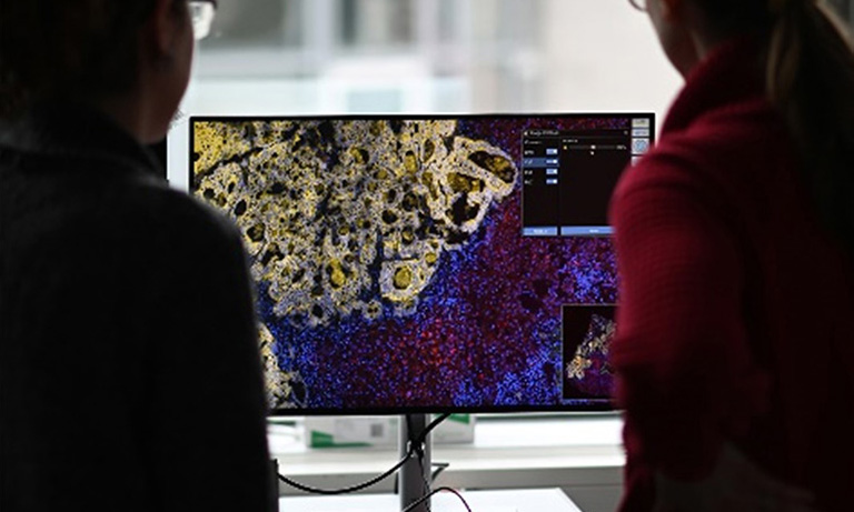
One of the biggest advantages of digitization is the ability to quickly review slides on a monitor, allowing us to analyze both broad overviews and detailed disease markers more efficiently. Digital pathology also facilitates genetic testing, which is often required to predict the effectiveness of molecular-targeted drug treatments for malignant tumors.
For accurate genetic testing, it’s essential that the test sample contains a sufficient number of tumor cells and is properly prepared. Digitized slides enable us to review and compare multiple images simultaneously, making it easier to select the most suitable samples for genetic testing.
As we outsource genetic testing, we can also share precise tumor area information with partner labs, improving both the accuracy and efficiency of genetic testing.
Q4. What is needed to expand digitalized operations?
We believe that reducing both the time and effort required for scanning, along with minimizing data storage requirements, is key to expanding digitized operations. One significant challenge is the cost associated with maintaining large-capacity storage servers.
If scanners could further reduce image file sizes without compromising quality, these improvements would facilitate broader adoption of whole slide scanning technology across veterinary medicine.

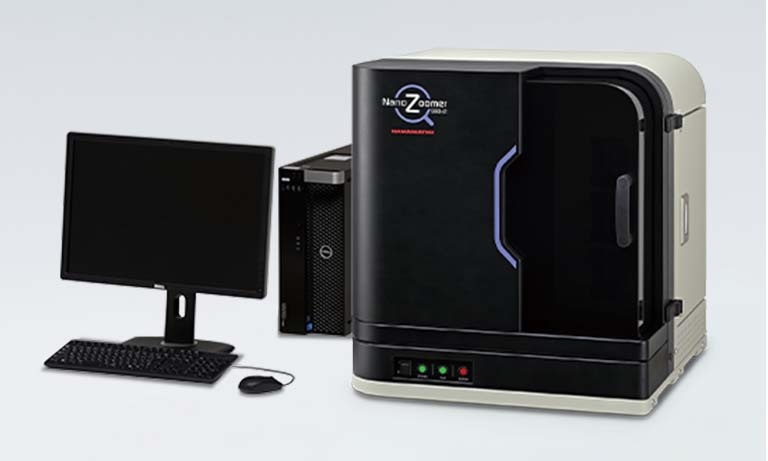
Q5: What are the benefits of NanoZoomer for your workflow?
When we used other vendors’ whole slide scanners, scanning was time-consuming, and we needed three scanners to process a large volume of samples within limited timeframes.
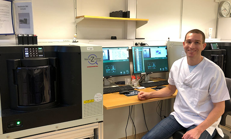
Since adopting the NanoZoomer in 2020, its fast scanning speed has enabled us to digitize the same number of samples with just one scanner. The scanning accuracy is also superior to previous models, significantly reducing the need for re-scanning. Currently, we only need to re-scan about 5 out of every 200 slides.
Furthermore, the NanoZoomer makes it easy to adjust focus positions during scanning, and the images produced have vivid colors that closely match actual samples. These benefits allow us to conduct tests efficiently without unnecessary delays.
Q6: What are your expectations for NanoZoomer in the future?
I hope AI technology will be utilized to further enhance image quality. For example, there are differences in how contrast appears between H&E staining and immunostaining, and focus can sometimes be inconsistent depending on conditions. If AI could automatically adjust contrast and optimize focus based on staining type, it would improve both the accuracy and speed of tests.
Additionally, when scanning large tissue samples that require multiple slides, significant effort is needed to stitch together the divided sections. It would be highly beneficial if AI could seamlessly process these images without manual intervention, streamlining our workflow.

Introduction of Amanecer Co., Ltd.
With the aspiration to contribute to the 'dawn' (Spanish: Amanecer) of veterinary medicine through pathological tests, Amanecer Co., Ltd. was established in 1996. As a commercial lab specializing in pet animals, we handle over 30 000 pathology samples annually from animal hospitals nationwide. Recognizing our social responsibility as a company supporting veterinary medicine, we have been digitizing pathology test results and distributing the 'Amanecer Annual' for over 15 years. We have also published breed-specific data collections such as 'Dog Pathology Guide' and 'Cat Pathology Guide'.
Since 2013, we have been using whole slide scanners to create digital slides for our test service. We also receive requests for data preparation for academic presentations and tasks for national and public research institutions. Now in our 29th year, we aim to continue sharing the awareness of “clinical pathology” with all our employees and clinical veterinarians, striving for higher standards of testing and better service provision.

President and CEO Dr. Hidetoshi Takahashi
Veterinarian, Doctor of Medicine (left)
Head of Testing Operations Department Dr. Kiyoshi Aita
Clinical Laboratory Technician, Doctor of Medicine (right)
*The content presented on this page reflects information available at the time of the interview.
The NanoZoomer line-up and medical device regulatory status varies across countries. For more information, please contact your local Hamamatsu sales representative.

Other research case study


- Confirmation
-
It looks like you're in the . If this is not your location, please select the correct region or country below.
You're headed to Hamamatsu Photonics website for GB (English). If you want to view an other country's site, the optimized information will be provided by selecting options below.
In order to use this website comfortably, we use cookies. For cookie details please see our cookie policy.
- Cookie Policy
-
This website or its third-party tools use cookies, which are necessary to its functioning and required to achieve the purposes illustrated in this cookie policy. By closing the cookie warning banner, scrolling the page, clicking a link or continuing to browse otherwise, you agree to the use of cookies.
Hamamatsu uses cookies in order to enhance your experience on our website and ensure that our website functions.
You can visit this page at any time to learn more about cookies, get the most up to date information on how we use cookies and manage your cookie settings. We will not use cookies for any purpose other than the ones stated, but please note that we reserve the right to update our cookies.
1. What are cookies?
For modern websites to work according to visitor’s expectations, they need to collect certain basic information about visitors. To do this, a site will create small text files which are placed on visitor’s devices (computer or mobile) - these files are known as cookies when you access a website. Cookies are used in order to make websites function and work efficiently. Cookies are uniquely assigned to each visitor and can only be read by a web server in the domain that issued the cookie to the visitor. Cookies cannot be used to run programs or deliver viruses to a visitor’s device.
Cookies do various jobs which make the visitor’s experience of the internet much smoother and more interactive. For instance, cookies are used to remember the visitor’s preferences on sites they visit often, to remember language preference and to help navigate between pages more efficiently. Much, though not all, of the data collected is anonymous, though some of it is designed to detect browsing patterns and approximate geographical location to improve the visitor experience.
Certain type of cookies may require the data subject’s consent before storing them on the computer.
2. What are the different types of cookies?
This website uses two types of cookies:
- First party cookies. For our website, the first party cookies are controlled and maintained by Hamamatsu. No other parties have access to these cookies.
- Third party cookies. These cookies are implemented by organizations outside Hamamatsu. We do not have access to the data in these cookies, but we use these cookies to improve the overall website experience.
3. How do we use cookies?
This website uses cookies for following purposes:
- Certain cookies are necessary for our website to function. These are strictly necessary cookies and are required to enable website access, support navigation or provide relevant content. These cookies direct you to the correct region or country, and support security and ecommerce. Strictly necessary cookies also enforce your privacy preferences. Without these strictly necessary cookies, much of our website will not function.
- Analytics cookies are used to track website usage. This data enables us to improve our website usability, performance and website administration. In our analytics cookies, we do not store any personal identifying information.
- Functionality cookies. These are used to recognize you when you return to our website. This enables us to personalize our content for you, greet you by name and remember your preferences (for example, your choice of language or region).
- These cookies record your visit to our website, the pages you have visited and the links you have followed. We will use this information to make our website and the advertising displayed on it more relevant to your interests. We may also share this information with third parties for this purpose.
Cookies help us help you. Through the use of cookies, we learn what is important to our visitors and we develop and enhance website content and functionality to support your experience. Much of our website can be accessed if cookies are disabled, however certain website functions may not work. And, we believe your current and future visits will be enhanced if cookies are enabled.
4. Which cookies do we use?
There are two ways to manage cookie preferences.
- You can set your cookie preferences on your device or in your browser.
- You can set your cookie preferences at the website level.
If you don’t want to receive cookies, you can modify your browser so that it notifies you when cookies are sent to it or you can refuse cookies altogether. You can also delete cookies that have already been set.
If you wish to restrict or block web browser cookies which are set on your device then you can do this through your browser settings; the Help function within your browser should tell you how. Alternatively, you may wish to visit www.aboutcookies.org, which contains comprehensive information on how to do this on a wide variety of desktop browsers.
5. What are Internet tags and how do we use them with cookies?
Occasionally, we may use internet tags (also known as action tags, single-pixel GIFs, clear GIFs, invisible GIFs and 1-by-1 GIFs) at this site and may deploy these tags/cookies through a third-party advertising partner or a web analytical service partner which may be located and store the respective information (including your IP-address) in a foreign country. These tags/cookies are placed on both online advertisements that bring users to this site and on different pages of this site. We use this technology to measure the visitors' responses to our sites and the effectiveness of our advertising campaigns (including how many times a page is opened and which information is consulted) as well as to evaluate your use of this website. The third-party partner or the web analytical service partner may be able to collect data about visitors to our and other sites because of these internet tags/cookies, may compose reports regarding the website’s activity for us and may provide further services which are related to the use of the website and the internet. They may provide such information to other parties if there is a legal requirement that they do so, or if they hire the other parties to process information on their behalf.
If you would like more information about web tags and cookies associated with on-line advertising or to opt-out of third-party collection of this information, please visit the Network Advertising Initiative website http://www.networkadvertising.org.
6. Analytics and Advertisement Cookies
We use third-party cookies (such as Google Analytics) to track visitors on our website, to get reports about how visitors use the website and to inform, optimize and serve ads based on someone's past visits to our website.
You may opt-out of Google Analytics cookies by the websites provided by Google:
https://tools.google.com/dlpage/gaoptout?hl=en
As provided in this Privacy Policy (Article 5), you can learn more about opt-out cookies by the website provided by Network Advertising Initiative:
http://www.networkadvertising.org
We inform you that in such case you will not be able to wholly use all functions of our website.
Close