Why NanoZoomer

Products

Solutions

Case study
Resources


United Kingdom (EN)
Select your region or country.
Beyond Standard Use: Unique Application of NanoZoomer® Scanning Technology
Dedicated to advancing applied science, Dr. Dirk Schaudien is a Veterinary Pathologist with a specialization in toxicopathology. He leads an experienced team offering comprehensive pathology services to industry clients, research institutions, and universities. The team brings deep expertise in toxicological pathology, tumor histopathology, and immunohistochemical characterization. Their work also includes conducting toxicity and carcinogenicity studies, as well as cell proliferation investigations, all in accordance with Good Laboratory Practice (GLP) standards.
Leveraging NanoZoomer® Scanners for High Throughput and Fluorescent Multiplexing
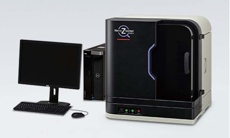
Since 2018, their state-of-the-art facilities include digital pathology scanners from Hamamatsu Photonics, expanding their research capabilities. Dr. Dirk Schaudien explains that the department invested in glass slide scanners to facilitate image analysis and documentation of special cases. Unlike standard clinical pathology departments, their lab does not have a continuous daily flow of samples.
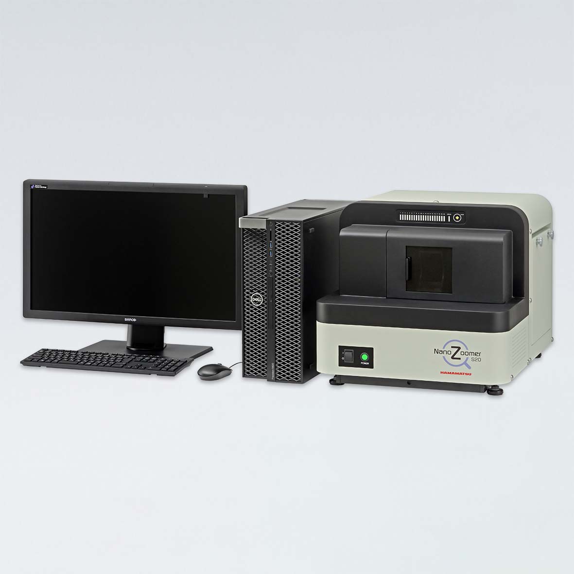
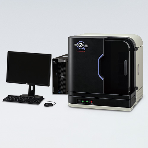
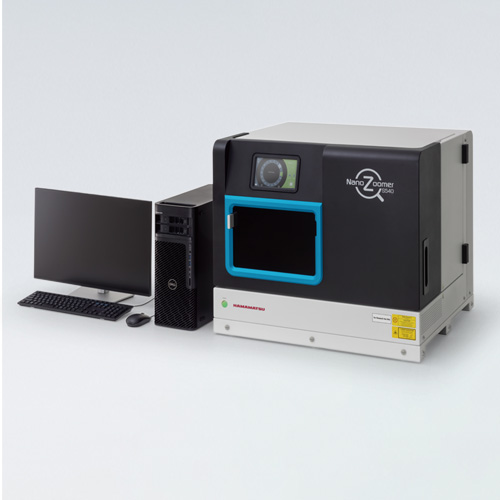
“We experience peaks in slide production associated with specific projects. Suddenly, we might have thousands of samples that need to be scanned,” he says. “In addition to handling brightfield (BF) samples, we also use fluorescent multiplexing for example to characterize different types of lymphocytes. That’s why we decided on two Hamamatsu scanners, the NanoZoomer S210 for its high capacity and throughput and the NanoZoomer S60 with its fluorescent unit. The S60 not only gives us additional capacity to scan BF samples during peak times but also allows us to scan fluorescent multiplex samples.”
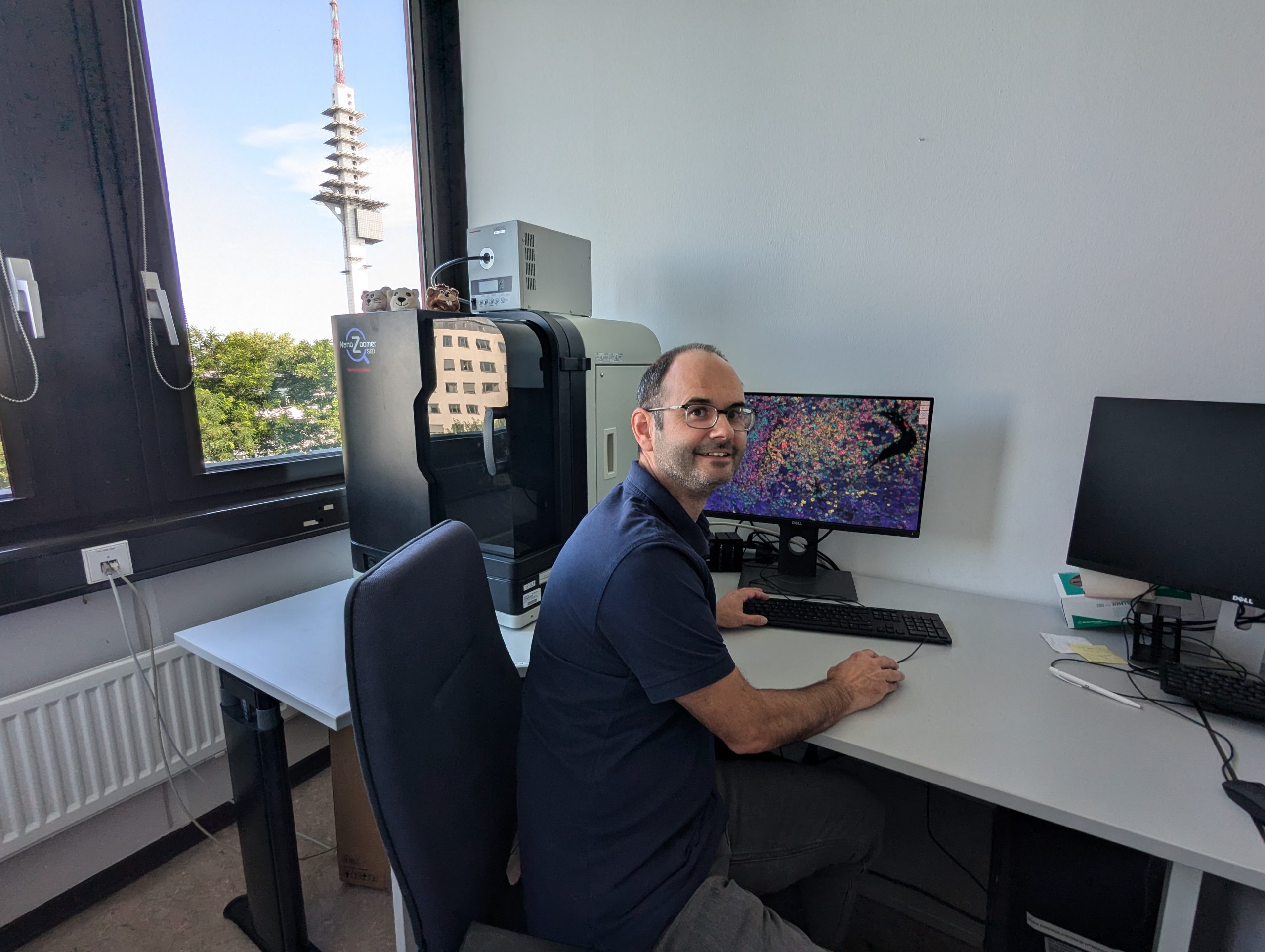
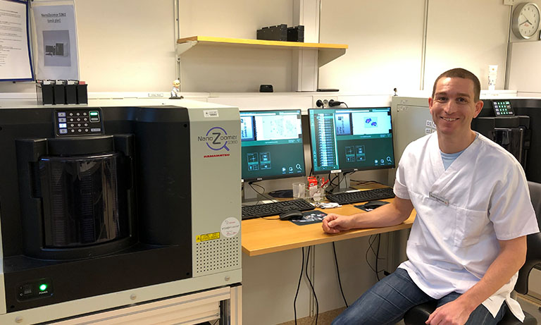
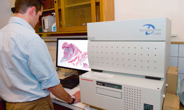
Dr. Dirk Schaudien with a NanoZoomer® S60.
Innovative Customization of the NanoZoomer® S60 for Enhanced Fluorescence Imaging
The Pathology team maximized the capabilities of their NanoZoomer S60, pushing it beyond standard usage. Collaborating with Hamamatsu’s application engineers, they developed a customized setup that allows the scanning of seven fluorescent channels on a single sample—surpassing the typical five or six channels. This was achieved thanks to the NanoZoomer S60’s flexible design, which accommodates multiple excitation and emission filter sets and beam splitters, allowing the team to expand the scanning capabilities beyond typical standards (see Fig. 1).
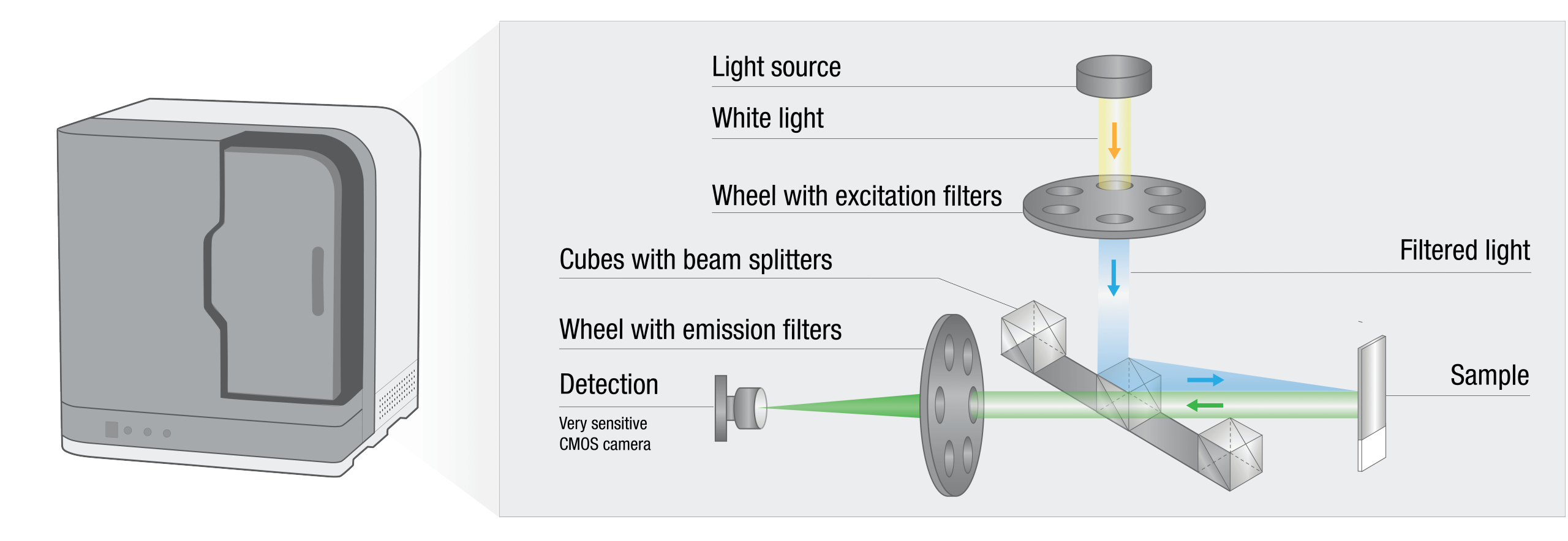
Fig. 1. Schematic representation of fluorescent scanning in the NanoZoomer® S60. The white light passes through an excitation filter, is reflected by a beam splitter, and is directed toward the sample. The fluorescent light emitted by the sample goes through the beam splitter and is filtered through the emission filter. It is then detected by a very sensitive CMOS camera. The NanoZoomer® S60 has two wheels, allowing the installation of six excitation and emission filters. The seventh hole in the emission wheel can be used when a single-band emission filter is installed on a cube. The NanoZoomer® S60 allows you to install three filter cubes.
Using the advanced capabilities of the NanoZoomer® S60, Dr. Schaudien's team explored the effects of low-frequency magnetic fields (ELF-MF) on the immune system in young mice, as part of research into childhood leukemia. The study focused on a mouse model mimicking a common genetic mutation associated with B-cell acute lymphoblastic leukemia (B-ALL), the most common form of childhood leukemia.
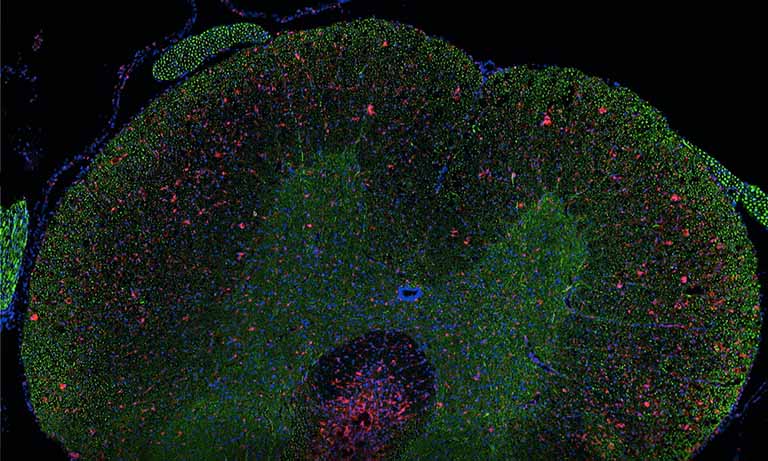
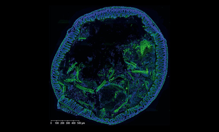
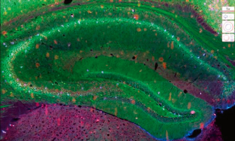
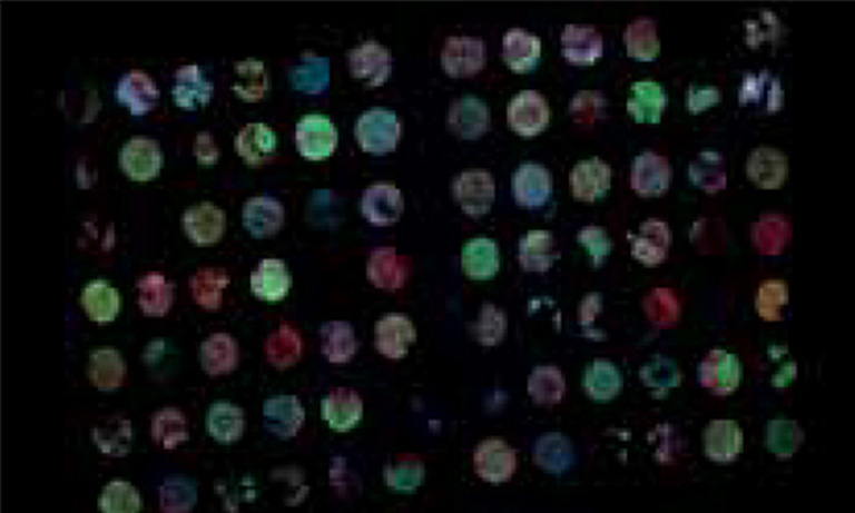
The seven-channel setup provided by the NanoZoomer S60 allowed the team to analyze how ELF-MF exposure, both before and after birth, impacted immune cells in tissues such as blood, spleen, bone marrow, and thymus. One example of their findings is a fluorescent image of the spleen captured using the S60 scanner, where six different immune system markers are stained in six distinct colors, with the seventh color representing the nuclear marker DAPI.
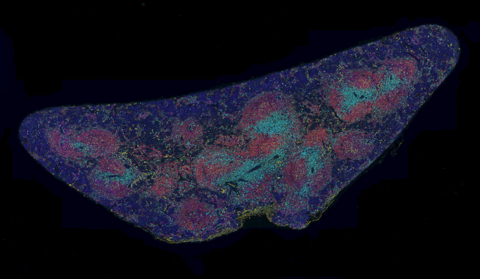
Mouse spleen with multiplex staining
Blue – Nuclei, cyan – CD4 positive cells, green – CD3 positive cells, yellow – immunoglobuline M (IgM), orange – CD19, red – CD45R positive cells, dark red – CD8 positive cells.
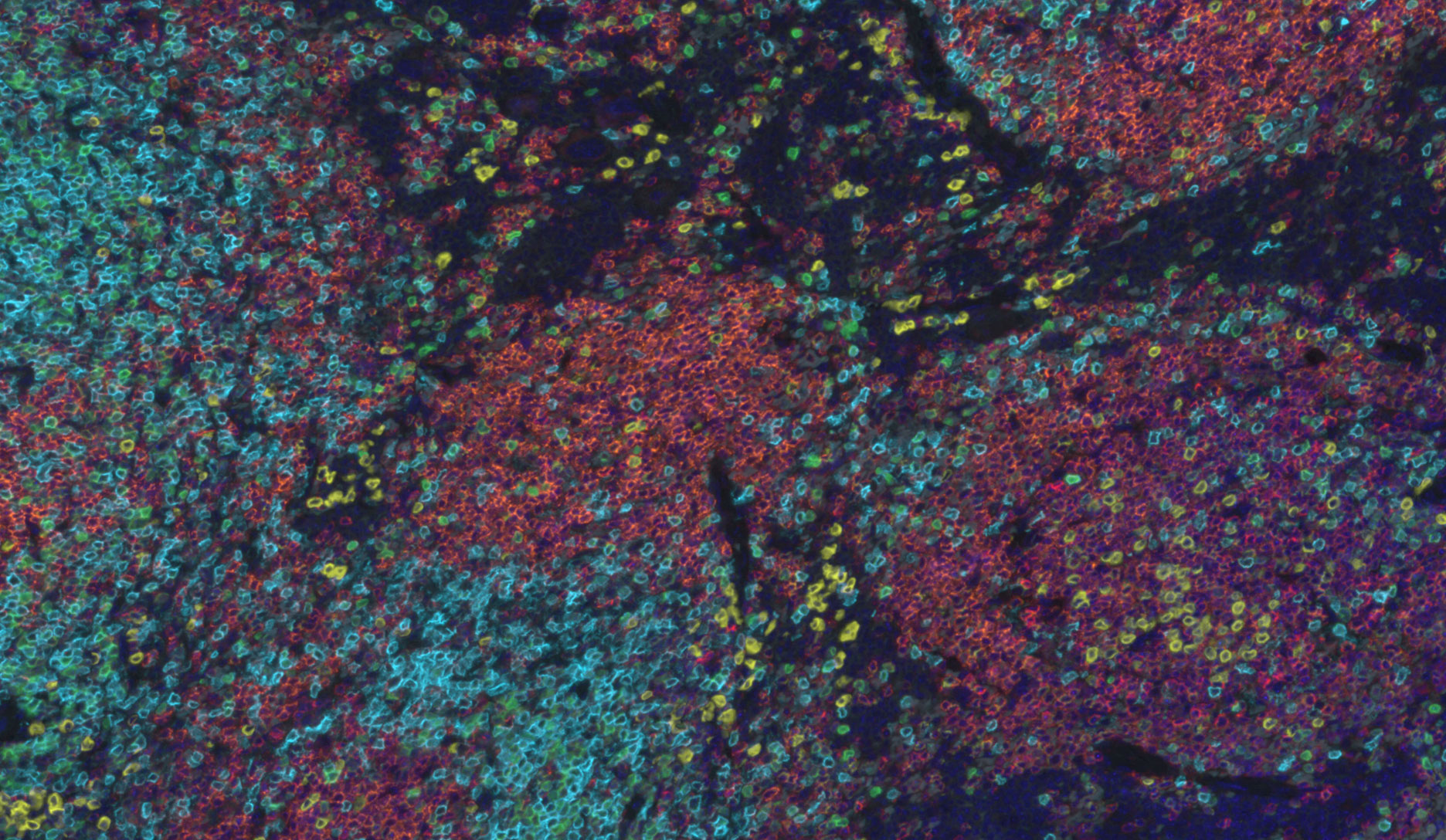
Merge 40x magnification
Blue – Nuclei, cyan – CD4 positive cells, green – CD3 positive cells, yellow – immunoglobuline M (IgM), orange – CD19, red – CD45R positive cells, dark red – CD8 positive cells.
| Fluorophore | Central Wavelength | Ex Filter | Em Filter | Beam splitter | Antigen |
|---|---|---|---|---|---|
| DAPI | Ex 359 Em 457 |
360/23 |
440/40 | Cube 1 | Nuclei |
| Discovery DCC kit (Roche) | Ex 436 Em 480 |
436/22 (Cube 3) | 480/20 | Cube 3 | CD4 |
| Discovery FAM kit (Roche) | Ex 490 Em 520 |
485/20 | 525/30 | Cube 1 | CD3 |
| Discovery Rhodamine 6G kit (Roche) | Ex 546 Em 572 |
535/26 (Cube 3) |
572/15 | Cube 3 | IgM |
| Discovery Red 610 kit (Roche) | Ex 580 Em 625 |
592/8 (Cube 2) |
628/32 (Cube 2) | Cube 2 | CD19 |
| Discovery Cy5 kit (Roche) | Ex 650 Em 670 |
650/13 | 684/24 | Cube 1 | CD45R |
| Opal Polaris 780 | Ex 768 Em 780 |
740/13 | 809/81 | Cube 1 | CD8 |
Table 1. List of fluorophores used in the study, including the specific excitation and emission filter sets that have been installed in the NanoZoomer® S60. Cube 1: pentaband beam splitter, Cube 2: a single band beam splitter mounted together with an optimal excitation and an emission filter to detect “Red 610”, Cube 3: a dual beamsplitter mounted together with a dual excitation filter.
Advancing Digital Pathology through collaborative Efforts
The collaboration with Hamamatsu Photonics demonstrates the impact of partnerships in digital pathology. This team effort has not only expanded the technological limits of fluorescent imaging but also opened new possibilities for scientific exploration, demonstrating the power of innovation through partnership.
Researcher profile

Dr. Dirk Schaudien is a Veterinary Pathologist with a specialization in toxicopathology. He is highly interested in image acquisition and analysis and likes to go beyond existing limitations to achieve better results.
About the author

Marta Böhle, PhD, is an Application Engineer at Hamamatsu, specializing in both research and clinical whole slide imaging using NanoZoomer® scanners. With a PhD in Neuroscience and extensive experience in diverse imaging techniques, Marta is passionate about imaging and dedicated to supporting customers in their projects.
Important notice: The NanoZoomer® S60 series includes IVD medical devices and research use only devices. Fluorescence option is available for research use only devices.
Other research case studies


- Confirmation
-
It looks like you're in the . If this is not your location, please select the correct region or country below.
You're headed to Hamamatsu Photonics website for GB (English). If you want to view an other country's site, the optimized information will be provided by selecting options below.
In order to use this website comfortably, we use cookies. For cookie details please see our cookie policy.
- Cookie Policy
-
This website or its third-party tools use cookies, which are necessary to its functioning and required to achieve the purposes illustrated in this cookie policy. By closing the cookie warning banner, scrolling the page, clicking a link or continuing to browse otherwise, you agree to the use of cookies.
Hamamatsu uses cookies in order to enhance your experience on our website and ensure that our website functions.
You can visit this page at any time to learn more about cookies, get the most up to date information on how we use cookies and manage your cookie settings. We will not use cookies for any purpose other than the ones stated, but please note that we reserve the right to update our cookies.
1. What are cookies?
For modern websites to work according to visitor’s expectations, they need to collect certain basic information about visitors. To do this, a site will create small text files which are placed on visitor’s devices (computer or mobile) - these files are known as cookies when you access a website. Cookies are used in order to make websites function and work efficiently. Cookies are uniquely assigned to each visitor and can only be read by a web server in the domain that issued the cookie to the visitor. Cookies cannot be used to run programs or deliver viruses to a visitor’s device.
Cookies do various jobs which make the visitor’s experience of the internet much smoother and more interactive. For instance, cookies are used to remember the visitor’s preferences on sites they visit often, to remember language preference and to help navigate between pages more efficiently. Much, though not all, of the data collected is anonymous, though some of it is designed to detect browsing patterns and approximate geographical location to improve the visitor experience.
Certain type of cookies may require the data subject’s consent before storing them on the computer.
2. What are the different types of cookies?
This website uses two types of cookies:
- First party cookies. For our website, the first party cookies are controlled and maintained by Hamamatsu. No other parties have access to these cookies.
- Third party cookies. These cookies are implemented by organizations outside Hamamatsu. We do not have access to the data in these cookies, but we use these cookies to improve the overall website experience.
3. How do we use cookies?
This website uses cookies for following purposes:
- Certain cookies are necessary for our website to function. These are strictly necessary cookies and are required to enable website access, support navigation or provide relevant content. These cookies direct you to the correct region or country, and support security and ecommerce. Strictly necessary cookies also enforce your privacy preferences. Without these strictly necessary cookies, much of our website will not function.
- Analytics cookies are used to track website usage. This data enables us to improve our website usability, performance and website administration. In our analytics cookies, we do not store any personal identifying information.
- Functionality cookies. These are used to recognize you when you return to our website. This enables us to personalize our content for you, greet you by name and remember your preferences (for example, your choice of language or region).
- These cookies record your visit to our website, the pages you have visited and the links you have followed. We will use this information to make our website and the advertising displayed on it more relevant to your interests. We may also share this information with third parties for this purpose.
Cookies help us help you. Through the use of cookies, we learn what is important to our visitors and we develop and enhance website content and functionality to support your experience. Much of our website can be accessed if cookies are disabled, however certain website functions may not work. And, we believe your current and future visits will be enhanced if cookies are enabled.
4. Which cookies do we use?
There are two ways to manage cookie preferences.
- You can set your cookie preferences on your device or in your browser.
- You can set your cookie preferences at the website level.
If you don’t want to receive cookies, you can modify your browser so that it notifies you when cookies are sent to it or you can refuse cookies altogether. You can also delete cookies that have already been set.
If you wish to restrict or block web browser cookies which are set on your device then you can do this through your browser settings; the Help function within your browser should tell you how. Alternatively, you may wish to visit www.aboutcookies.org, which contains comprehensive information on how to do this on a wide variety of desktop browsers.
5. What are Internet tags and how do we use them with cookies?
Occasionally, we may use internet tags (also known as action tags, single-pixel GIFs, clear GIFs, invisible GIFs and 1-by-1 GIFs) at this site and may deploy these tags/cookies through a third-party advertising partner or a web analytical service partner which may be located and store the respective information (including your IP-address) in a foreign country. These tags/cookies are placed on both online advertisements that bring users to this site and on different pages of this site. We use this technology to measure the visitors' responses to our sites and the effectiveness of our advertising campaigns (including how many times a page is opened and which information is consulted) as well as to evaluate your use of this website. The third-party partner or the web analytical service partner may be able to collect data about visitors to our and other sites because of these internet tags/cookies, may compose reports regarding the website’s activity for us and may provide further services which are related to the use of the website and the internet. They may provide such information to other parties if there is a legal requirement that they do so, or if they hire the other parties to process information on their behalf.
If you would like more information about web tags and cookies associated with on-line advertising or to opt-out of third-party collection of this information, please visit the Network Advertising Initiative website http://www.networkadvertising.org.
6. Analytics and Advertisement Cookies
We use third-party cookies (such as Google Analytics) to track visitors on our website, to get reports about how visitors use the website and to inform, optimize and serve ads based on someone's past visits to our website.
You may opt-out of Google Analytics cookies by the websites provided by Google:
https://tools.google.com/dlpage/gaoptout?hl=en
As provided in this Privacy Policy (Article 5), you can learn more about opt-out cookies by the website provided by Network Advertising Initiative:
http://www.networkadvertising.org
We inform you that in such case you will not be able to wholly use all functions of our website.
Close