Why NanoZoomer

Products

Solutions

Case study
Resources


United States (EN)
Select your region or country.
Benefits of Digital Pathology and the importance of slide preparation
Digital Pathology at a multisite Swedish laboratory: Region Skåne
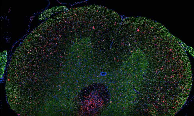
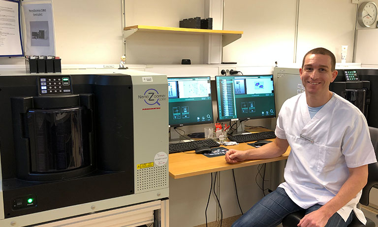
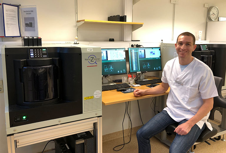
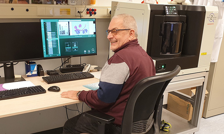
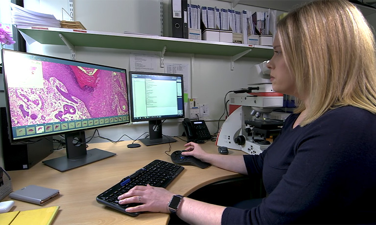
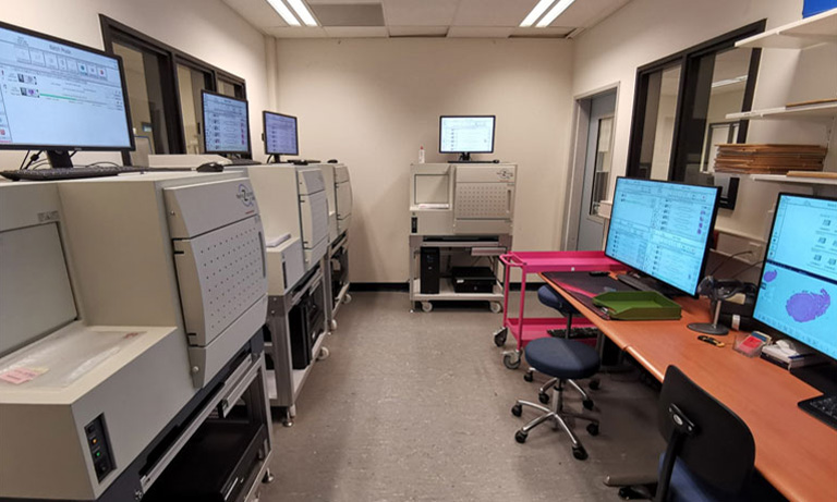
In recent years, digital pathology has become increasingly recognised as a clinical diagnostic tool and has taken a crucial role in pathology laboratories. One of the early adopters of digital pathology is the Department of Clinical Genetics and Pathology, Office for Medical Services, Region Skåne, a multisite Swedish laboratory which produces over 800,000 histopathology slides per year. The department has implemented digital pathology in all four labs across its organisation, making it one of the world’s largest digital pathology groups in clinical practice. In total, 18 digital slide scanners are installed, including a combination of NanoZoomerⓇ S360 Digital slide scanner and NanoZoomer S60 Digital slide scanner.

Leading up to 2019, it was recognised that there was a shortage of reporting capacity within the current pathologist team. There was also a need to reduce cancer care lead time. Digital pathology was identified as an option to solve both of these shortfalls. The site in Malmö was active in the implementation and integration of digital pathology that ran throughout 2019 and involved all laboratories. From November 2019 onwards the region was 100% digital with regards to histopathology. The implementation was an intensive process. Clear and structured feedback was required from pathologists to assess any concerns to enable the laboratory to react and make any necessary corrections.
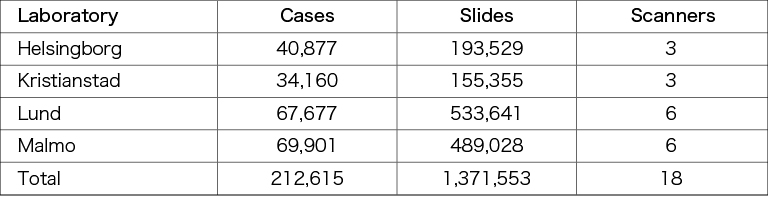
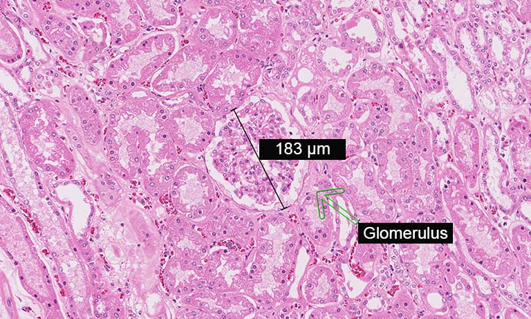
Table 1. Total amount of cases and slides produced by the Department of Clinical Genetics and Pathology, Region Skåne from May 2019 until the end of 2020. All slides are scanned for digital pathology.
Dr. Kevin Sandeman, Clinical Pathologist and Head of Unit for the pathologists in Malmö, shares his experiences and learnings from their successful implementation of digital pathology. He explains the advantages that digital pathology has brought to the laboratory and discusses the importance of standardised slide preparation.
Dr. Kevin Sandeman, Clinical Pathologist and Head of Unit for the pathologists in the Department of Clinical Genetics and Pathology, Malmö.
What are the advantages of digital pathology?
Dr. Sandeman comments, “Whilst there are many advantages to digital pathology, being able to work remotely, particularly during a global pandemic such as COVID-19, is a huge advantage to the region. It also allows group consultation to be carried out without face-to-face contact.
Faster diagnosis and, in turn, a reduction of cancer care lead times has also been seen across the region. Faster diagnosis is a key indicator and evidence shows that the introduction of digital pathology has reduced the diagnostic proportion of our reporting times by up to 27% with a decrease in total time to diagnosis of 12.5 %.”
From a financial stand point the implementation of digital pathology has resulted in cost savings by minimising the amount of pathologist overtime required by the region.
“Faster diagnosis is a key indicator and evidence shows that the introduction of digital pathology has reduced the
diagnostic proportion of our reporting times by up to 27% with a decrease in total time to diagnosis of 12.5 %.”
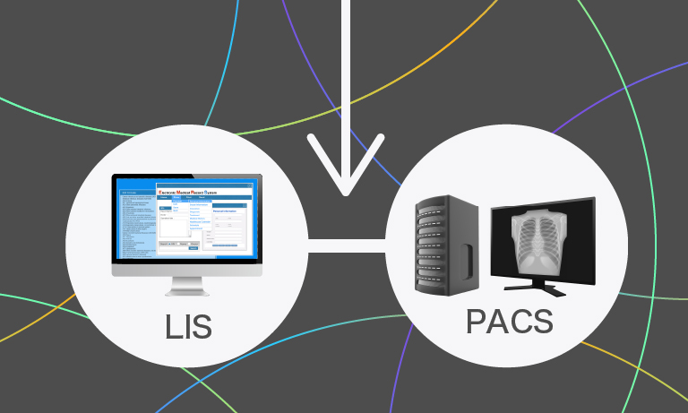
Standardisation of the laboratory processes throughout the region has allowed them to redistribute wet specimens before preparation. This enables laboratories who previously may have struggled to complete their work to send specimens to nearby laboratories and be assured their specimens will be treated in the same way. The scanned images are then still available for all users through the central image management system.
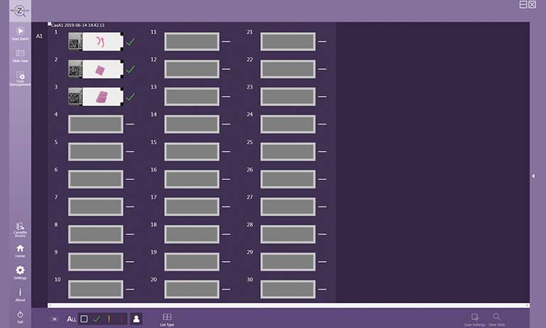
“The NanoZoomer scanners used by the Skåne region are set up to scan the majority of tissue and stains under a single profile.” This creates a smooth workflow with minimal interaction from the laboratory technicians. Dr. Sandeman reports that the training of resident pathologists is now more streamlined with the advantage of communication via annotations on the images. It also allows the specialist pathologist to have an overview of all the training cases.
Has the introduction of digital pathology affected the laboratory workflow?
“Slide preparation is a key factor in the success of digital pathology (…).”
Dr. Sandeman explains, “The laboratory workflow has remained largely the same. Training of staff using the scanner is important and helps them realise the importance of how the laboratory process affects the final scanned image. Standardising the laboratory process as a whole has also eased the implementation of digital pathology.” Slide preparation is a key factor in the success of digital pathology and the laboratory has seen an increase in the quality of work produced since its introduction.
Standardisation and quality of pre-analytical procedures
For digital pathology to meet its full potential, labs have to focus on establishing quality and standardisation throughout all pre-analytical processes.
Dr. Sandeman highlights that “transitioning to digital is a hard quality check for the laboratory”, as the quality of the digital image completely depends on the quality of the tissue slide that is scanned. Slide preparation, therefore, is key to achieving high-quality digital images. Variation in pre-analytical processes leads to variation in colour and image quality. As all these pre-analytical procedures influence the final scanned image, standardisation makes visualisation and interpretation of the digital image easier.
Dr. Sandeman adds that variation in slide preparation and handling between centres can result in conflicts in scanning results. Thus, technicians of all centres have to standardise pre-analytical procedures. An example of this is to standardise the thickness of sections in their microtomy step which streamlines the scanning process. Communication between centres for standardised solutions was an important factor in successfully implementing digital pathology.
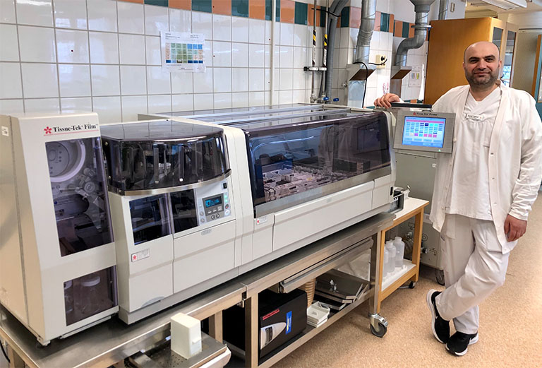
Technician Walid Jabbar next to the Tissue-Tek Prisma® Plus and Tissue-Tek Film® integrated tissue staining and film coverslipping system.
Facilitating the scanning process
It is also important for the laboratory to optimise pre-analytical factors to facilitate the scanning process. In particular, the choice of coverslips can have great influence on this. All centres of region Skåne employ both glass and film coverslippers (Tissue-Tek Film® coverslipper, Sakura Finetek).

“The quality of digital images is similar for both film and glass coverslipped slides.”
In total, eight Tissue-Tek Film® coverslippers are installed. Dr. Sandeman reports that the quality of the digital images is similar for both film and glass coverslipped slides. The technicians that prepare the slides found that coverslipping with film facilitates the scanning process and minimises out-of-focus events. Though routine stained slides are coverslipped with film coverslips as well as glass coverslips, they experience many advantages when using the film coverslipper:
- The coverslipping process with film is faster compared to glass coverslipping
- Slides coverslipped with film have a shorter drying time than glass coverslipped slides. The coverslipped slides can be stacked without sticking together and are immediately ready for scanning
- The faster scanning process and short drying time provides a seamless synchronisation with high-throughput scanners
- There are no mounting media spills when coverslipping with film that might interfere with the scanner and disrupt the scanning process.
“Film coverslipping is well suited [to digital pathology] in terms of the scanning process”
Dr. Sandeman emphasises that the technicians in Malmö have a clear preference for the film coverslippers. He adds that with film coverslipping, the coverslip is positioned centrally and in a same way for every slide, eliminating the manual adjustment that may occur if a glass coverslip is not aligned properly to the slide.
The future
The rise of new advanced technologies is pushing the field of digital pathology even further. One specific technology that is expected to be integrated increasingly into digital pathology is artificial intelligence (AI). Dr. Sandeman is an independent researcher in the field of AI. When asked about how important standardisation of slide preparation is in digital pathology, he states that “standardised preparation of slides is central in AI”. He explains further that AI algorithms are trained for a specific task, therefore every artefact will have to be taken into consideration when training the algorithm. “If you do not want to constantly update the algorithm intended for a specific task, standardisation of the slide preparation is important.”
The NanoZoomer line-up and medical device regulatory status varies across countries. For more information, please contact your local Hamamatsu sales representative.

Other clinical case study


- Confirmation
-
It looks like you're in the . If this is not your location, please select the correct region or country below.
You're headed to Hamamatsu Photonics website for US (English). If you want to view an other country's site, the optimized information will be provided by selecting options below.
In order to use this website comfortably, we use cookies. For cookie details please see our cookie policy.
- Cookie Policy
-
This website or its third-party tools use cookies, which are necessary to its functioning and required to achieve the purposes illustrated in this cookie policy. By closing the cookie warning banner, scrolling the page, clicking a link or continuing to browse otherwise, you agree to the use of cookies.
Hamamatsu uses cookies in order to enhance your experience on our website and ensure that our website functions.
You can visit this page at any time to learn more about cookies, get the most up to date information on how we use cookies and manage your cookie settings. We will not use cookies for any purpose other than the ones stated, but please note that we reserve the right to update our cookies.
1. What are cookies?
For modern websites to work according to visitor’s expectations, they need to collect certain basic information about visitors. To do this, a site will create small text files which are placed on visitor’s devices (computer or mobile) - these files are known as cookies when you access a website. Cookies are used in order to make websites function and work efficiently. Cookies are uniquely assigned to each visitor and can only be read by a web server in the domain that issued the cookie to the visitor. Cookies cannot be used to run programs or deliver viruses to a visitor’s device.
Cookies do various jobs which make the visitor’s experience of the internet much smoother and more interactive. For instance, cookies are used to remember the visitor’s preferences on sites they visit often, to remember language preference and to help navigate between pages more efficiently. Much, though not all, of the data collected is anonymous, though some of it is designed to detect browsing patterns and approximate geographical location to improve the visitor experience.
Certain type of cookies may require the data subject’s consent before storing them on the computer.
2. What are the different types of cookies?
This website uses two types of cookies:
- First party cookies. For our website, the first party cookies are controlled and maintained by Hamamatsu. No other parties have access to these cookies.
- Third party cookies. These cookies are implemented by organizations outside Hamamatsu. We do not have access to the data in these cookies, but we use these cookies to improve the overall website experience.
3. How do we use cookies?
This website uses cookies for following purposes:
- Certain cookies are necessary for our website to function. These are strictly necessary cookies and are required to enable website access, support navigation or provide relevant content. These cookies direct you to the correct region or country, and support security and ecommerce. Strictly necessary cookies also enforce your privacy preferences. Without these strictly necessary cookies, much of our website will not function.
- Analytics cookies are used to track website usage. This data enables us to improve our website usability, performance and website administration. In our analytics cookies, we do not store any personal identifying information.
- Functionality cookies. These are used to recognize you when you return to our website. This enables us to personalize our content for you, greet you by name and remember your preferences (for example, your choice of language or region).
- These cookies record your visit to our website, the pages you have visited and the links you have followed. We will use this information to make our website and the advertising displayed on it more relevant to your interests. We may also share this information with third parties for this purpose.
Cookies help us help you. Through the use of cookies, we learn what is important to our visitors and we develop and enhance website content and functionality to support your experience. Much of our website can be accessed if cookies are disabled, however certain website functions may not work. And, we believe your current and future visits will be enhanced if cookies are enabled.
4. Which cookies do we use?
There are two ways to manage cookie preferences.
- You can set your cookie preferences on your device or in your browser.
- You can set your cookie preferences at the website level.
If you don’t want to receive cookies, you can modify your browser so that it notifies you when cookies are sent to it or you can refuse cookies altogether. You can also delete cookies that have already been set.
If you wish to restrict or block web browser cookies which are set on your device then you can do this through your browser settings; the Help function within your browser should tell you how. Alternatively, you may wish to visit www.aboutcookies.org, which contains comprehensive information on how to do this on a wide variety of desktop browsers.
5. What are Internet tags and how do we use them with cookies?
Occasionally, we may use internet tags (also known as action tags, single-pixel GIFs, clear GIFs, invisible GIFs and 1-by-1 GIFs) at this site and may deploy these tags/cookies through a third-party advertising partner or a web analytical service partner which may be located and store the respective information (including your IP-address) in a foreign country. These tags/cookies are placed on both online advertisements that bring users to this site and on different pages of this site. We use this technology to measure the visitors' responses to our sites and the effectiveness of our advertising campaigns (including how many times a page is opened and which information is consulted) as well as to evaluate your use of this website. The third-party partner or the web analytical service partner may be able to collect data about visitors to our and other sites because of these internet tags/cookies, may compose reports regarding the website’s activity for us and may provide further services which are related to the use of the website and the internet. They may provide such information to other parties if there is a legal requirement that they do so, or if they hire the other parties to process information on their behalf.
If you would like more information about web tags and cookies associated with on-line advertising or to opt-out of third-party collection of this information, please visit the Network Advertising Initiative website http://www.networkadvertising.org.
6. Analytics and Advertisement Cookies
We use third-party cookies (such as Google Analytics) to track visitors on our website, to get reports about how visitors use the website and to inform, optimize and serve ads based on someone's past visits to our website.
You may opt-out of Google Analytics cookies by the websites provided by Google:
https://tools.google.com/dlpage/gaoptout?hl=en
As provided in this Privacy Policy (Article 5), you can learn more about opt-out cookies by the website provided by Network Advertising Initiative:
http://www.networkadvertising.org
We inform you that in such case you will not be able to wholly use all functions of our website.
Close