Why NanoZoomer

Products

Solutions

Case study
Resources


United States (EN)
Select your region or country.
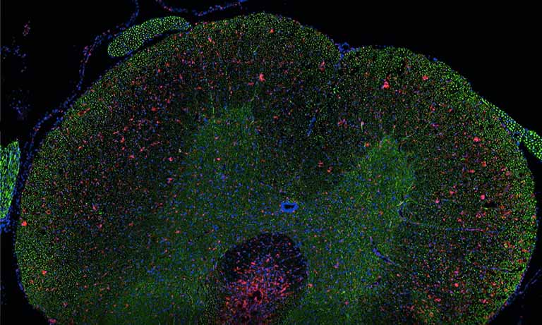
And still it scans! 15 years of experience with Hamamatsu NanoZoomer
Erasmus Medical Center Rotterdam
We recently had the pleasure of interviewing Dr. Peter Riegman from Erasmus University. Dr. Riegman is a Molecular Biologist and head of the Erasmus MC Tissue Bank. As an early adopter of digital pathology, Dr. Riegman purchased his first Hamamatsu Photonics whole slide scanner in 2005. It was a NanoZoomerⓇ-HT, one of Hamamatsu’s very first production units, and it is still in daily use at Riegman’s pathology lab today. It sits side by side with another NanoZoomer-HT installed in 2009 and a NanoZoomer S360 Digital slide scanner installed in 2019. This trifecta of NZ scanners image 350 slides per day, with the capacity to scan up to 700 slides per day.

Dr. Riegman believes the image quality and service are the two critical elements for successful digital pathology implementation.
Quality flexible solutions
Under Dr. Riegman’s guidance, the lab automated slide scanning to comply with routine diagnostic procedures. The lab also provides special stains as a service and this created a unique challenge that needed to be addressed. Through collaboration, Hamamatsu provided a small adjustment to the scanner that enabled Erasmus to distinguish the slide lab labels from the special stain barcode. This relatively minor tuning was critical for fully automated slide scanning and this flexibility within the NanoZoomer design allowed for broader adoption within Erasmus University. Dr. Riegman’s experience with Hamamatsu NanoZoomers illustrates the commitment Hamamatsu has to providing uncompromising digital pathology solutions that provide excellent image quality along with reliability and intelligent, flexible design.

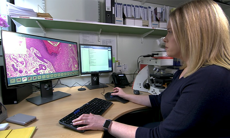
Dr. Peter Riegman, Head of the Erasmus MC Tissue Bank
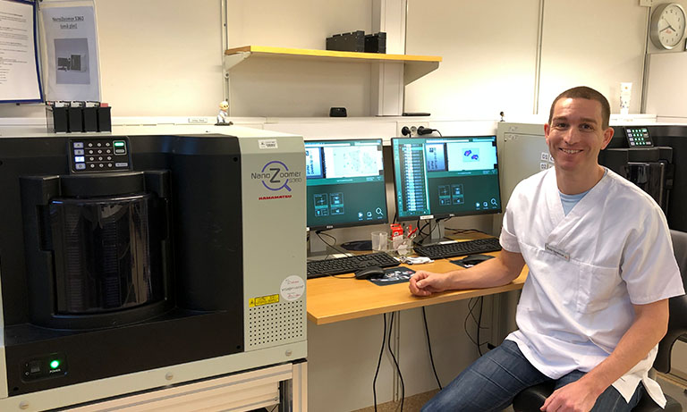
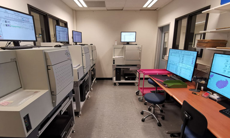
The scanner room
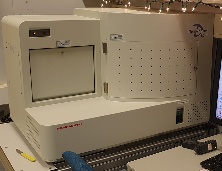
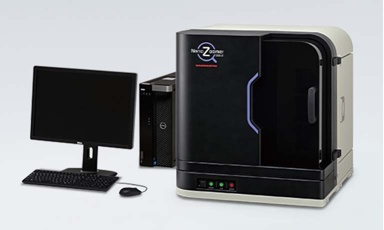
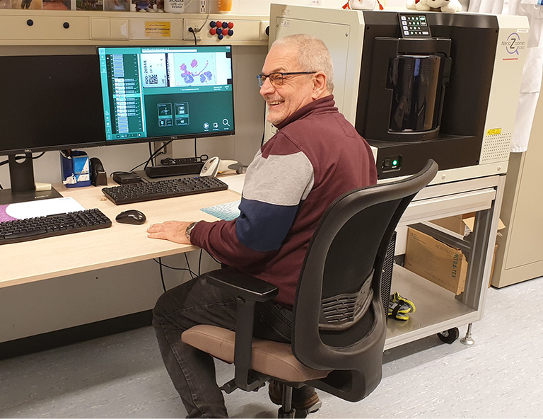
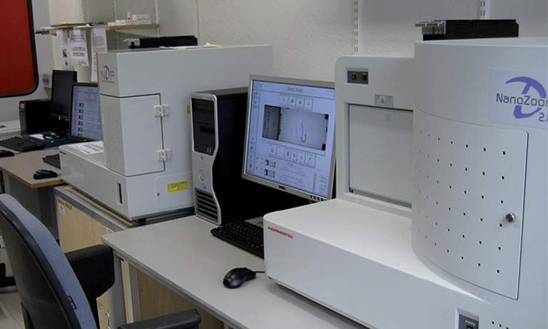
NanoZoomer 2.0-HT
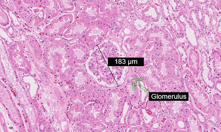
Dr. Riegman had the opportunity to compare the image quality of both NanoZoomer-HT and NanoZoomer S360 Digital slide scanner. He concludes the image quality of NanoZoomer-HT and NanoZoomer S360 Digital slide scanner are sufficient for routine diagnosis.
Reliability
A successful digital pathology implementation starts with organizations addressing the unmet needs and reimaging the laboratory workflow to increase efficiencies. At the time of purchase, the emphasis is often on image quality, ease of use, and costs. Upfront costs are easy to understand, but the actual return on investment comes from long-term reliability that maintains consistent slide throughput increasing lab efficiency.
Erasmus MC Tissue Bank operates two older scanners, so how reliable are those scanners?
These scanners do not require a lot of maintenance, contrary to what people may think. At times unexpected things do happen, such as replacing the light unit or filters after many years of overuse. In general, the correction step in NanoZoomer-HT scanner offset the issues, and the Hamamatsu service engineers can troubleshoot and fix those without noteworthy disruption to the workflow.
Hamamatsu NanoZoomer
Hamamatsu NanoZoomer scanners are built on the foundation and knowledge of over15 years of product innovation in digital pathology combined with 65 years of photonics experience. Hamamatsu believes in manufacturing and providing the most reliable whole slide scanners in the market. We thrive on product and image quality.
Visit nanozoomer.com to find out more about Hamamatsu NanoZoomer scanners.
Please enjoy the full interview with Dr. Riegman:
Dr. Peter Riegman, Head of the Erasmus MC Tissue Bank, was one of the first NanoZoomer customers worldwide. Actually, the third ever-built NanoZoomer was delivered in 2005 to the Erasmus Institute. When the workload became too much for one scanner, a second NanoZoomer-HT was installed in 2009 and in 2019, an NanoZoomer S360 Digital slide scanner was added.
You currently have three scanners: two (very old) NanoZoomer-HT and one NanoZoomer S360 Digital slide scanner. Both NanoZoomer-HTs are still scanning – do they still work properly and do you need a lot of maintenance in the meantime?
Not a lot of maintenance, although sometimes unexpected things need to be replaced. For example, the light unit or filters need to be taken out due to the fact the glass fibers that send the light to the stage is worn out and the light changes slightly in color over the many years. This is of no influence to the image quality, because this is corrected by the NanoZoomer-HT, but the effect finally became out of range for this correction. In addition, computers need to be updated to Windows 10. One done and one to go.
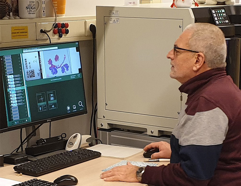
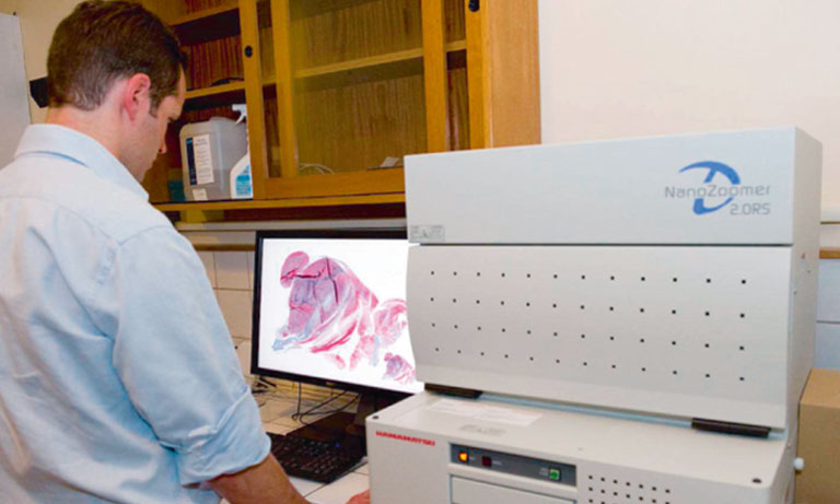
Dr. Riegman at image quality inspection
After such a long time, it was to be expected, that the system had to be updated to a new Windows version. Did you face difficulties with the Windows 10 upgrade? Especially with the old NanoZoomer-HTs?
After installation of Windows 10 on the Windows 7 computer, we asked Hamamatsu for an installable version of the scan software. The question alone was enough for them to completely install everything remotely for us. It works flawlessly since that time.
The repairs do not sound like standard maintenance issues. Did Hamamatsu find the failures quickly?
Yes, the support engineers dealt with failures swiftly as with the repairs.
Things like degrading fibers are surely not the standard repair issues?
The repairs were certainly not standard issues; however, Hamamatsu support asked for the log files, drew the conclusion, and knew what to do.
You have been using the NanoZoomer S360 Digital slide scanner for a year now. How does it compare to the old NanoZoomer-HTs?
The high image quality of the NanoZoomer S360 Digital slide scanner is comparable with the fine quality of the both NanoZoomer-HTs. Moreover, for the average diagnostics the images are fine.
Of course, speed of the NanoZoomer S360 Digital slide scanner is so much faster due to new techniques and much improved logistics in the scanner.
In the past, you used the NanoZoomer-HT for research, routine and – as one of the first to digitize their training slides – students’ education. Is that still the main use?
In the past the slow scanners were used for science, education and limited diagnostic applications* like external consulting, panels and molecular diagnostic archive for glass slides needed to be sacrificed for molecular assessments.
The NanoZoomer S360 Digital slide scanner is now scanning for diagnostics in general on a daily basis. This way we run tests to enable us to scale up to scan the total yearly production of 250.000 glass slides.
And the NanoZoomer-HTs? Are they still used for the same applications?
Yes, we use the NanoZoomer-HTs still for the old applications.
Do you plan to go digital even for routine diagnostics?
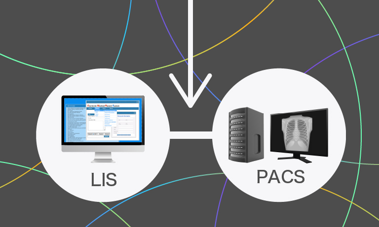
Yes. The next step is that we want to add a modern pathology communication program or IMS that makes the scanners communicate with the LIMS so the pathologists are supported in their digital workflow and after that, we scale up the scanning capacity to scan the full production.
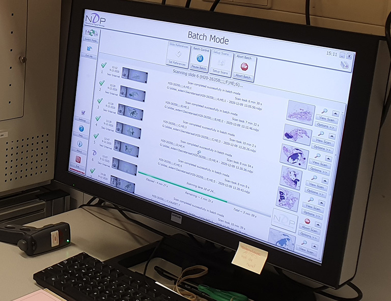
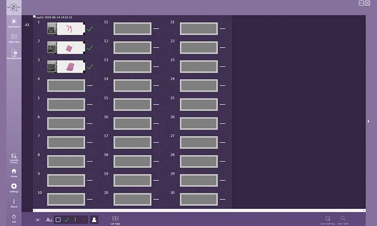
Batch processing
In the beginning, you scanned most of your slides in semiautomatic mode, as the scanner software accidentally identified felt tip pen marks as tissue and therefore did not define the scan area correctly, leading to longer scan times and larger file sizes. With the fast NanoZoomer S360 Digital slide scanner, and if you plan to scan your whole production in future, did you find a way to scan all slides in fully automatic mode?
Yes. Especially, for routine diagnostics it is a pre-requisite even to scan fully automatically. We optimized the laboratory for the preparation of slides and logistics, like the labeling of the slides with bar codes. Automatic staining covering and drying of the slides was adjusted. Some adjustments were made on the scanner to distinguish the lab labels from the special staining equipment bar codes that can also be present on a glass slide.
Back in 2005 after installation of the first scanner, its integration in the workflow took time and some modifications by Hamamatsu. Do you still face sometimes problems with compatibility?
We do seem to have a good working logistics in scanning for diagnostic purposes in place and good insight in the needed communication. We now need a communication program or IMS to make the logistics complete to enhance the pathologists’ acceptance level.
Are there functions or features, you would like Hamamatsu to improve or to add?
For the scanner itself it would help if direct access to empty slots for cassettes could be given during scanning without bothering the ongoing process. For instance, a place/chamber where new cassettes could be put in (e.g. max four cassettes at a time) which are added by the scanner to the carousel whenever the scanning process allows that. When the cassettes enter the carousel, inventory is made and priority can be given. In addition, a place/chamber where the scanner puts finished cassettes (e.g. max 4 cassettes at a time) when all slides in the cassette are scanned to satisfaction. This way you create a sort of running belt and decrease personnel waiting and operating time.
* Note: NanoZoomer S360 Digital slide scanner is CE marked under EU’s In Vitro Diagnostics Directive (IVDD) for in vitro diagnostic use.
NanoZoomer S360 Digital slide scanner is for Research Use Only in US. For other countries, please consult with Hamamatsu. NanoZoomer-HT is for Research Use Only.
The NanoZoomer line-up and medical device regulatory status varies across countries. For more information, please contact your local Hamamatsu sales representative.

Other clinical case study


- Confirmation
-
It looks like you're in the . If this is not your location, please select the correct region or country below.
You're headed to Hamamatsu Photonics website for US (English). If you want to view an other country's site, the optimized information will be provided by selecting options below.
In order to use this website comfortably, we use cookies. For cookie details please see our cookie policy.
- Cookie Policy
-
This website or its third-party tools use cookies, which are necessary to its functioning and required to achieve the purposes illustrated in this cookie policy. By closing the cookie warning banner, scrolling the page, clicking a link or continuing to browse otherwise, you agree to the use of cookies.
Hamamatsu uses cookies in order to enhance your experience on our website and ensure that our website functions.
You can visit this page at any time to learn more about cookies, get the most up to date information on how we use cookies and manage your cookie settings. We will not use cookies for any purpose other than the ones stated, but please note that we reserve the right to update our cookies.
1. What are cookies?
For modern websites to work according to visitor’s expectations, they need to collect certain basic information about visitors. To do this, a site will create small text files which are placed on visitor’s devices (computer or mobile) - these files are known as cookies when you access a website. Cookies are used in order to make websites function and work efficiently. Cookies are uniquely assigned to each visitor and can only be read by a web server in the domain that issued the cookie to the visitor. Cookies cannot be used to run programs or deliver viruses to a visitor’s device.
Cookies do various jobs which make the visitor’s experience of the internet much smoother and more interactive. For instance, cookies are used to remember the visitor’s preferences on sites they visit often, to remember language preference and to help navigate between pages more efficiently. Much, though not all, of the data collected is anonymous, though some of it is designed to detect browsing patterns and approximate geographical location to improve the visitor experience.
Certain type of cookies may require the data subject’s consent before storing them on the computer.
2. What are the different types of cookies?
This website uses two types of cookies:
- First party cookies. For our website, the first party cookies are controlled and maintained by Hamamatsu. No other parties have access to these cookies.
- Third party cookies. These cookies are implemented by organizations outside Hamamatsu. We do not have access to the data in these cookies, but we use these cookies to improve the overall website experience.
3. How do we use cookies?
This website uses cookies for following purposes:
- Certain cookies are necessary for our website to function. These are strictly necessary cookies and are required to enable website access, support navigation or provide relevant content. These cookies direct you to the correct region or country, and support security and ecommerce. Strictly necessary cookies also enforce your privacy preferences. Without these strictly necessary cookies, much of our website will not function.
- Analytics cookies are used to track website usage. This data enables us to improve our website usability, performance and website administration. In our analytics cookies, we do not store any personal identifying information.
- Functionality cookies. These are used to recognize you when you return to our website. This enables us to personalize our content for you, greet you by name and remember your preferences (for example, your choice of language or region).
- These cookies record your visit to our website, the pages you have visited and the links you have followed. We will use this information to make our website and the advertising displayed on it more relevant to your interests. We may also share this information with third parties for this purpose.
Cookies help us help you. Through the use of cookies, we learn what is important to our visitors and we develop and enhance website content and functionality to support your experience. Much of our website can be accessed if cookies are disabled, however certain website functions may not work. And, we believe your current and future visits will be enhanced if cookies are enabled.
4. Which cookies do we use?
There are two ways to manage cookie preferences.
- You can set your cookie preferences on your device or in your browser.
- You can set your cookie preferences at the website level.
If you don’t want to receive cookies, you can modify your browser so that it notifies you when cookies are sent to it or you can refuse cookies altogether. You can also delete cookies that have already been set.
If you wish to restrict or block web browser cookies which are set on your device then you can do this through your browser settings; the Help function within your browser should tell you how. Alternatively, you may wish to visit www.aboutcookies.org, which contains comprehensive information on how to do this on a wide variety of desktop browsers.
5. What are Internet tags and how do we use them with cookies?
Occasionally, we may use internet tags (also known as action tags, single-pixel GIFs, clear GIFs, invisible GIFs and 1-by-1 GIFs) at this site and may deploy these tags/cookies through a third-party advertising partner or a web analytical service partner which may be located and store the respective information (including your IP-address) in a foreign country. These tags/cookies are placed on both online advertisements that bring users to this site and on different pages of this site. We use this technology to measure the visitors' responses to our sites and the effectiveness of our advertising campaigns (including how many times a page is opened and which information is consulted) as well as to evaluate your use of this website. The third-party partner or the web analytical service partner may be able to collect data about visitors to our and other sites because of these internet tags/cookies, may compose reports regarding the website’s activity for us and may provide further services which are related to the use of the website and the internet. They may provide such information to other parties if there is a legal requirement that they do so, or if they hire the other parties to process information on their behalf.
If you would like more information about web tags and cookies associated with on-line advertising or to opt-out of third-party collection of this information, please visit the Network Advertising Initiative website http://www.networkadvertising.org.
6. Analytics and Advertisement Cookies
We use third-party cookies (such as Google Analytics) to track visitors on our website, to get reports about how visitors use the website and to inform, optimize and serve ads based on someone's past visits to our website.
You may opt-out of Google Analytics cookies by the websites provided by Google:
https://tools.google.com/dlpage/gaoptout?hl=en
As provided in this Privacy Policy (Article 5), you can learn more about opt-out cookies by the website provided by Network Advertising Initiative:
http://www.networkadvertising.org
We inform you that in such case you will not be able to wholly use all functions of our website.
Close