Why NanoZoomer

Products
Solutions

Case study
Resources


United States (EN)
Select your region or country.
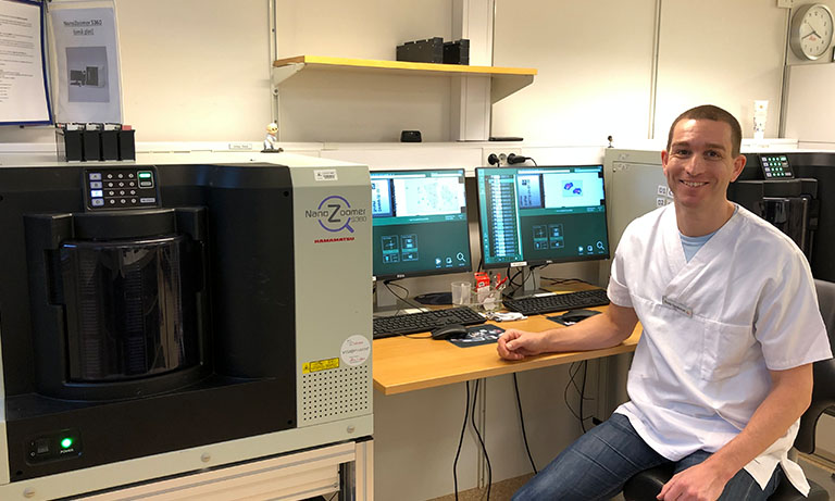
Erasmus MC Tissue Bank, their long-term experience using NanoZoomer HT
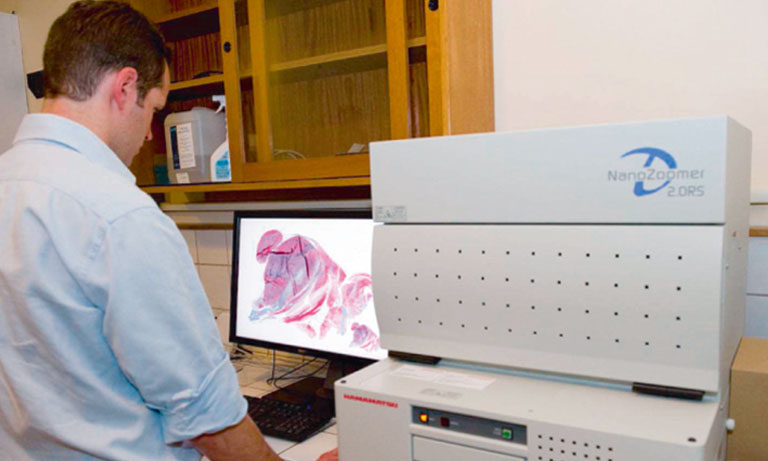
The advantage of virtual microscopy is that virtual slides are, in contrast to their glass counterparts, easily accessible from remote locations when stored on an image server. Like glass slides under a microscope they can be viewed with different magnifications, pathologists can navigate through the tissue and add annotations to mark important areas. In contrast to images taken with a standard camera mounted on a microscope, a virtual slide enables the pathologist to review the whole tissue and not only the small section imaged by the camera. This is particularly important for second opinion revisions. Furthermore virtual slides don’t fade with time, like their glass counterparts do. Fluorescence labelled tissues are especially prone to bleaching, making it very difficult to review the original slides after weeks or even days after preparation.
Dr. Peter Riegman, Head of the Erasmus MC Tissue Bank, was one of the first NanoZoomer customers worldwide. Actually the 3rd ever built NanoZoomer was delivered in 2005 to the Erasmus Institute and has been used there ever since.
“Our initial idea was to establish a Tissue Micro Array platform. Slides are scanned and afterwards they can be analysed by dedicated software and algorithms” Dr. Riegman tells us. “But of course our hope was that it could be used for pathology purposes, too.”
“It turns out that he was correct: the TMA platform was started only two years ago. The NanoZoomer is used basically for three application fields”, explains Dr. Bas de Jong, Assistant Manager of the Erasmus MC Tissue Bank. Namely for:
- research (imaging facility for internal and external scientists, digital analysis)
- diagnostics (virtual revision slides for second opinion, internal and for other hospitals, case studies)
- students' education
It runs nearly full time (i.e. 24 h, 7 days/week) with a throughput of ca.700 slides/week. In the past three years no slides have been broken by the scanner. In the very beginning it sometimes happened that slides were not handled correctly, owing mostly to a cover slip or label sticker being placed askew. But thanks to regular feedback from Dr. Riegman and his colleagues, Hamamatsu have continued to improve both the software and hardware, making the NanoZoomer the most reliable system on the market.
Dr. Bas de Jong estimates that the only maintenance needed in the past 5 years have been to exchange the light bulb in the NanoZoomer, which was done by himself and his co-workers, without needing to call a service visit from a Hamamatsu support engineer.
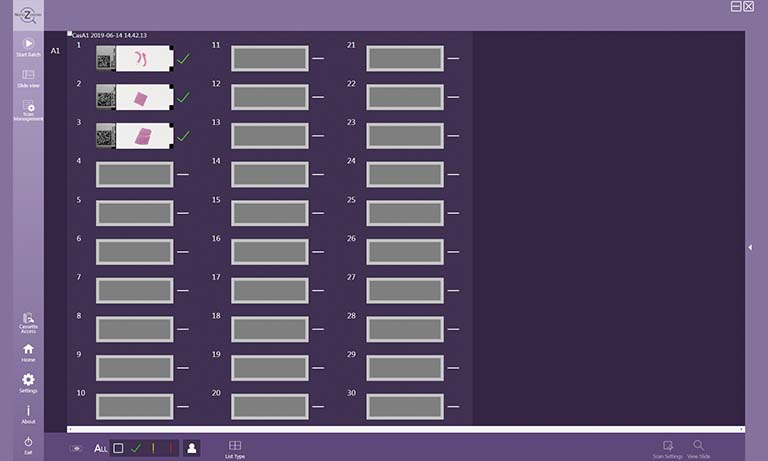
The NanoZoomer is designed to scan slides automatically, including tissue recognition, definition of the proper scan area and the scanning itself. This automatic mode works even with slides marked with felt tip notes.
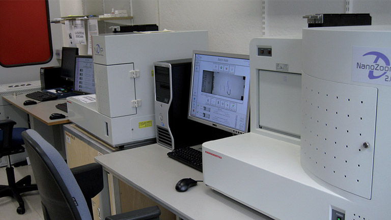
Yet in the experience of Dr. de Jong and Dr. Riegman, felt tip pens exhibit similar contrast as stained tissue in the black and white macro image, consequently the scan area sometimes is defined too large, leading to longer than necessary scanning times and larger file size. For this reason they scan 99% of their slides in semi-automatic mode. This mode allows the operator to review the scan area and focus before scanning. The NanoZoomer is also capable of setting different scan properties for different slides. This functionality enables the operator to scan very diverse slides in batches rather than individually.
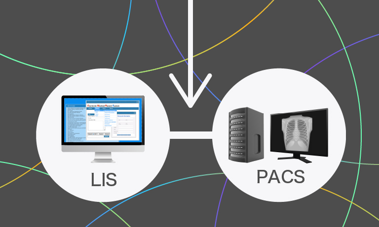
A short time ago Dr. Riegman and Dr. de Jong decided to upgrade the old scanner to version 2.0 to achieve higher throughput. But as the demand for virtual slides for research applications had increased so much, that even the upgraded scanner is used to capacity, a second scanner was purchased. “This scanner is connected to an image server and used for diagnostics, scanning ca. 50 slides per day. These slides are directly stored on the server, making network bandwidth a crucial factor for scanning speed” states Dr. de Jong. In his opinion the greatest challenge of using two of the fastest scanners on the market is storage capacity for handling the large amounts of image data.

Erasmus MC Tissue Bank, their long-term experience using NanoZoomer [1.4 MB/PDF]
* Note: NanoZoomer HT is for Research Use Only.
The NanoZoomer line-up and medical device regulatory status varies across countries. For more information, please contact your local Hamamatsu sales representative.

- Confirmation
-
It looks like you're in the . If this is not your location, please select the correct region or country below.
You're headed to Hamamatsu Photonics website for US (English). If you want to view an other country's site, the optimized information will be provided by selecting options below.
In order to use this website comfortably, we use cookies. For cookie details please see our cookie policy.
- Cookie Policy
-
This website or its third-party tools use cookies, which are necessary to its functioning and required to achieve the purposes illustrated in this cookie policy. By closing the cookie warning banner, scrolling the page, clicking a link or continuing to browse otherwise, you agree to the use of cookies.
Hamamatsu uses cookies in order to enhance your experience on our website and ensure that our website functions.
You can visit this page at any time to learn more about cookies, get the most up to date information on how we use cookies and manage your cookie settings. We will not use cookies for any purpose other than the ones stated, but please note that we reserve the right to update our cookies.
1. What are cookies?
For modern websites to work according to visitor’s expectations, they need to collect certain basic information about visitors. To do this, a site will create small text files which are placed on visitor’s devices (computer or mobile) - these files are known as cookies when you access a website. Cookies are used in order to make websites function and work efficiently. Cookies are uniquely assigned to each visitor and can only be read by a web server in the domain that issued the cookie to the visitor. Cookies cannot be used to run programs or deliver viruses to a visitor’s device.
Cookies do various jobs which make the visitor’s experience of the internet much smoother and more interactive. For instance, cookies are used to remember the visitor’s preferences on sites they visit often, to remember language preference and to help navigate between pages more efficiently. Much, though not all, of the data collected is anonymous, though some of it is designed to detect browsing patterns and approximate geographical location to improve the visitor experience.
Certain type of cookies may require the data subject’s consent before storing them on the computer.
2. What are the different types of cookies?
This website uses two types of cookies:
- First party cookies. For our website, the first party cookies are controlled and maintained by Hamamatsu. No other parties have access to these cookies.
- Third party cookies. These cookies are implemented by organizations outside Hamamatsu. We do not have access to the data in these cookies, but we use these cookies to improve the overall website experience.
3. How do we use cookies?
This website uses cookies for following purposes:
- Certain cookies are necessary for our website to function. These are strictly necessary cookies and are required to enable website access, support navigation or provide relevant content. These cookies direct you to the correct region or country, and support security and ecommerce. Strictly necessary cookies also enforce your privacy preferences. Without these strictly necessary cookies, much of our website will not function.
- Analytics cookies are used to track website usage. This data enables us to improve our website usability, performance and website administration. In our analytics cookies, we do not store any personal identifying information.
- Functionality cookies. These are used to recognize you when you return to our website. This enables us to personalize our content for you, greet you by name and remember your preferences (for example, your choice of language or region).
- These cookies record your visit to our website, the pages you have visited and the links you have followed. We will use this information to make our website and the advertising displayed on it more relevant to your interests. We may also share this information with third parties for this purpose.
Cookies help us help you. Through the use of cookies, we learn what is important to our visitors and we develop and enhance website content and functionality to support your experience. Much of our website can be accessed if cookies are disabled, however certain website functions may not work. And, we believe your current and future visits will be enhanced if cookies are enabled.
4. Which cookies do we use?
There are two ways to manage cookie preferences.
- You can set your cookie preferences on your device or in your browser.
- You can set your cookie preferences at the website level.
If you don’t want to receive cookies, you can modify your browser so that it notifies you when cookies are sent to it or you can refuse cookies altogether. You can also delete cookies that have already been set.
If you wish to restrict or block web browser cookies which are set on your device then you can do this through your browser settings; the Help function within your browser should tell you how. Alternatively, you may wish to visit www.aboutcookies.org, which contains comprehensive information on how to do this on a wide variety of desktop browsers.
5. What are Internet tags and how do we use them with cookies?
Occasionally, we may use internet tags (also known as action tags, single-pixel GIFs, clear GIFs, invisible GIFs and 1-by-1 GIFs) at this site and may deploy these tags/cookies through a third-party advertising partner or a web analytical service partner which may be located and store the respective information (including your IP-address) in a foreign country. These tags/cookies are placed on both online advertisements that bring users to this site and on different pages of this site. We use this technology to measure the visitors' responses to our sites and the effectiveness of our advertising campaigns (including how many times a page is opened and which information is consulted) as well as to evaluate your use of this website. The third-party partner or the web analytical service partner may be able to collect data about visitors to our and other sites because of these internet tags/cookies, may compose reports regarding the website’s activity for us and may provide further services which are related to the use of the website and the internet. They may provide such information to other parties if there is a legal requirement that they do so, or if they hire the other parties to process information on their behalf.
If you would like more information about web tags and cookies associated with on-line advertising or to opt-out of third-party collection of this information, please visit the Network Advertising Initiative website http://www.networkadvertising.org.
6. Analytics and Advertisement Cookies
We use third-party cookies (such as Google Analytics) to track visitors on our website, to get reports about how visitors use the website and to inform, optimize and serve ads based on someone's past visits to our website.
You may opt-out of Google Analytics cookies by the websites provided by Google:
https://tools.google.com/dlpage/gaoptout?hl=en
As provided in this Privacy Policy (Article 5), you can learn more about opt-out cookies by the website provided by Network Advertising Initiative:
http://www.networkadvertising.org
We inform you that in such case you will not be able to wholly use all functions of our website.
Close